Computational human phantoms are invaluable for calculating the dose to radiosensitive organs and tissues. However, the determined doses are only strictly accurate for a person with exactly the same anatomy as the phantom.
To improve this situation, researchers in Brazil have developed a series of 18 anthropometric adult human phantoms covering a wide range of body masses and heights. These are described in an article in the July 7, 2011, issue of Physics in Medicine and Biology (Vol. 56:13, pp. 3749-3772).
"Radiation doses received by a patient during radiological examinations depend, among other factors, on the anthropometric properties of the patient, especially their body mass," explained co-author Richard Kramer, PhD, of the Federal University of Pernambuco's department of nuclear energy. "Software packages based on reference phantoms cannot cover the variety of patient anatomies usually found in radiology. Therefore, we created phantoms that cover the range of body masses and standing heights applicable for most patients."
Phantom creation
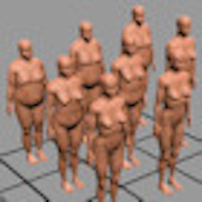
The researchers used anthropometric data to alter two mesh-based adult standing phantoms: the male MASH3 and female FASH3. Nine new phantoms were developed for each sex, based on 10th, 50th, and 90th mass and height percentiles of Caucasian populations averaged over nine countries.
 |
| Female anthropometric mesh phantoms for 10th, 50th, and 90th mass and height percentiles. Image courtesy of Physics in Medicine and Biology. |
Parameters for body mass and standing height were extracted from a visual anthropometry software package (PeopleSize, Open Ergonomics). Phantoms were developed assuming constant body mass index (BMI) for a given mass percentile and for different heights. For a given height, the change in body mass was considered to reflect mainly changes in subcutaneous adipose tissue (not organ masses). Organ mass was, however, scaled as a function of height, using information extracted from autopsy data.
The new phantoms were created as follows: First, the body masses and heights of the base phantoms MASH3 and FASH3 were increased to match the average 50th percentiles (65 kg and 163.8 cm for the female; 79 kg and 176.4 cm for the male). The adipose tissue levels in these "central" phantoms were then altered to model the 10th and 90th percentiles of mass. For the 10th percentile phantoms, muscle and/or soft tissue was also reduced if required.
Next, phantoms for the 10th and 90th height percentiles were modeled by adapting the central phantoms to the required heights. After scaling of organs according to autopsy data, volumes of soft, muscle, and adipose tissue were increased or decreased to meet the targeted body mass. Finally, these phantoms were also modeled for the 10th and 90th mass percentiles.
Dosimetry details
The researchers then examined how body mass and height influence dosimetric results. To achieve this, the 18 mesh phantoms were voxelized and connected to the EGSnrc Monte Carlo code. The team calculated organ and tissue equivalent doses for external whole-body exposure to various photon beams and internal exposure to gamma emitters.
When irradiated externally, adipose tissue attenuates the incident radiation, thus reducing the equivalent dose to tissues located below it. Adipose tissue, however, also gives rise to scatter, which increases the equivalent dose. Thus the resulting equivalent dose is a combination of the two effects, and is dependent upon the size and location of an organ or tissue relative to the adipose layer.
For organs located below the subcutaneous adipose tissue layer, such as the liver, colon, and stomach, equivalent dose per air kerma was seen to decrease with increasing body mass, with the level of reduction dependent on the incident photon energy. For example, for 10-MeV photons, the equivalent dose per air kerma to the colon wall decreased by 5.5% with increasing body mass.
For 20-keV diagnostic x-rays, the equivalent dose per air kerma decreased by a factor of 10.4 as body mass increased. The authors pointed out, though, that in x-ray diagnosis, absolute equivalent dose usually increases with larger body mass due to the need to increase mAs to maintain image quality.
For organs located at the surface, such as the breasts, testes, and skin, weak attenuation and increased backscatter caused the equivalent dose per air kerma to increase or remain constant with increasing body mass. For all external exposures, changes in height had little effect on the equivalent doses.
Internal exposure
The researchers also calculated specific absorbed fractions (SAFs) for the 18 phantoms exposed to gamma emitters concentrated in selected organs. Results showed that SAFs decreased with increasing height and increased with increasing body mass.
For example, with the lungs as the source, SAFs in the neighboring liver decreased by up to 12.7% with increasing height, due to the increase in interorgan distances and organ masses. With increasing mass, SAFs increased by up to 5.3%, due to radiation scatter in the larger amount of adipose tissue.
With the liver as the target organ and the bladder contents as the source, SAFs decreased by up to 32.8% with increasing height, the larger effect being due to the greater distance between target and source organs. The increase in SAFs with increasing body mass was also greater (up to 16.8%) due to increased scatter radiation caused by more adipose tissue between the target and source.
Kramer noted that the new anthropometric phantoms are "patient-related." In other words, if a patient has the same body mass and height of one of the phantoms, then they would receive a similar, but not necessarily identical, radiation dose.
As such, the phantoms are not suitable for radiotherapy treatment planning, where a "patient-specific" phantom is required instead. "This is a 3D anatomical model of a specific patient, which could be made using our modeling method in combination with medical images (CT or MR) of that specific patient," Kramer told MedicalPhysicsWeb.
© IOP Publishing Limited. Republished with permission from medicalphysicsweb, a community website covering fundamental research and emerging technologies in medical imaging and radiation therapy.