University of Michigan researchers said they saved a baby's life with the aid of a customized tracheal splint manufactured on a 3D printer using image data from a CT scan, in a case reported in the New England Journal of Medicine.
Kaiba Gionfriddo was just 20 months old when a collapsed bronchus threatened his life, and doctors were uncertain if he would leave the hospital alive, according to otolaryngologist Dr. Glenn Green and Scott Hollister, PhD, professor of biomedical and mechanical engineering at the University of Michigan.
The doctors obtained emergency clearance from the U.S. Food and Drug Administration (FDA) to create and implant a tracheal splint made from a biopolymer known as polycaprolactone. In February 2012, at C.S. Mott Children's Hospital, the splint was sewn around the patient's bronchus to open it and provide a kind of skeleton to support its growth. Over about a three-year period, the splint will be absorbed by the body.
"It was amazing. As soon as the splint was put in, the lungs started going up and down for the first time and we knew he was going to be OK," Green said in a statement.
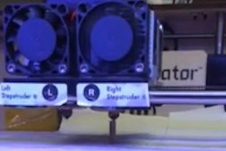
The research team crafted the device using high-resolution imaging and computer-aided design applied to a CT scan of the patient's trachea and bronchus, in a process that integrated an image-based computer model with laser-based 3D printing to produce the splint.
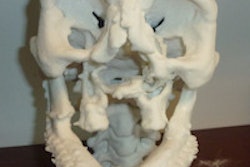
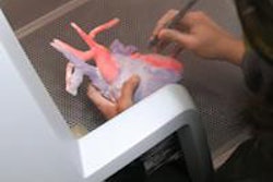
The CT-based design and 3D biomaterial printing process can be used to reconstruct a broad range of tissue structures. In preclinical models, for example, Green and Hollister have used the method to build ear and nose structures, as well as rebuild bone structures.