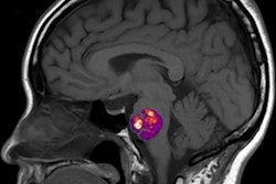
An artificial intelligence (AI)-based system developed to mimic how a radiologist interprets images can achieve performance comparable to that of academic neuroradiologists in diagnosing 19 common and rare diseases on brain MRI exams, according to research published online April 7 in Radiology.
The system combines deep learning-based detection of brain lesions, non-AI-based analysis of quantitative and clinical image features, and Bayesian networks in order to provide a differential diagnosis. In testing, it outperformed general radiologists and trainees and achieved results approaching that of neuroradiologists. It was created by a team of researchers led by Dr. Jeffrey Rudie, PhD, and Dr. Andreas Rauschecker, PhD, of the University of California, San Francisco.
Their AI system features three separate components. First, a convolutional neural network detects lesions based on abnormal fluid-attenuated inversion-recovery (FLAIR) signals from MRI scans. Next, image processing techniques are utilized to derive human-interpretable quantitative image and lesion features.
After these parameters are combined with five clinical features, a Bayesian network -- a probabilistic model of relationships between multiple variables -- then combines those quantitative features with five clinical features based on expert knowledge to arrive at a differential diagnosis.
"The AI system was constructed to separately model the perceptual and cognitive components of human radiologic image interpretation," the authors wrote.
The researcher compared the system's performance on a test set of 92 cases with that of four radiology residents, two neuroradiology fellows, two general radiologists, and two academic neuroradiology attending physicians.
| AI performance for diagnosing 19 brain diseases on MRI | |||||
| Radiology residents | General radiologists | Neuroradiology fellows | Academic neuroradiologists | AI system | |
| Mean accuracy* | 56% | 57% | 77% | 86% | 91% |
| Area under the curve | 0.73 | 0.72 | 0.85 | 0.90 | 0.92 |
"AI system performance was similar to that of academic neuroradiologists on all measures, was better than one of two neuroradiology fellows on top one differential diagnosis and top two differential diagnoses performance, and was consistently better than that of all radiology residents and general radiologists," they wrote.
The researchers noted that the radiologists across all specialization levels were better at diagnosing common diseases than they were at detecting rare diseases.
It's important now to test the AI system prospectively and at other institutions, according to the researchers. They expect, though, that it will generalize well, as the system has shown that it works across more than 20 different MR scanner types and a wide range of acquisition parameters.
"We anticipate that the general framework of a composite AI system combining data-driven and knowledge-driven approaches can be applied to many domains within radiology and will ultimately form the basis of more efficient and accurate radiology practice," they wrote.
In an accompanying editorial, Dr. Greg Zaharchuk, PhD, of Stanford University shared two thoughts on the research.
"One is the incredible amount of work that went into designing this AI system and its good performance on a narrow task, for which the authors should be congratulated," he wrote. "The other is how long the road ahead is before such a tool would be a must-have in the reading room, given the vast number of diseases that radiologists must recognize. It is possible that this methodological approach does not scale well with more added features and diseases."
However, this study "charts a plausible way forward to an automated diagnosis system that combines many AI methods guided by the domain expertise and training methods of human radiologists," Zaharchuck concluded.