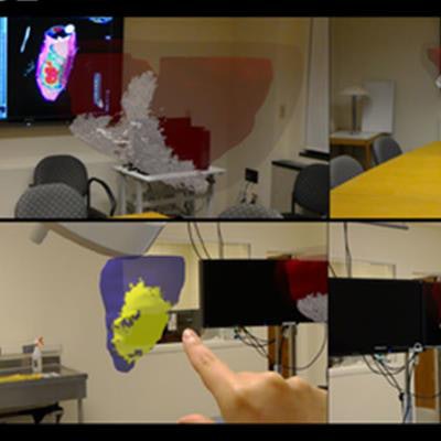
Augmented reality (AR) could someday allow clinicians to visualize 3D cardiac MRI scans while performing radiofrequency ablation to treat heart rhythm disorders, according to an article recently published online in PLOS One.
The group from Harvard Medical School used an AR headset (HoloLens, Microsoft) to examine 3D virtual heart models of five pigs in real-time as the animals underwent a simulated cardiac electrophysiological intervention. Looking at and interacting with these detailed 3D heart images -- based on MRI scans -- helped enhance the researchers' understanding of relevant anatomy during the procedure (PLOS One, October 8, 2018).
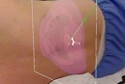
 Visualization of 3D pig heart models based on late gadolinium enhancement (LGE) cardiac MRI. Image courtesy of Jang et al.
On a follow-up questionnaire, the clinicians who performed the operation claimed that incorporating AR into surgical workflow would likely increase their effectiveness and performance overall.
"Physicians can now use AR to view 3D cardiac MR information with a touchless interaction in sterile environment," first author and doctoral candidate Jihye Jang said in a statement. "Our report shows exciting potential that having this complex 3D scar information through augmented reality during the intervention may help guide treatment and ultimately improve patient care."