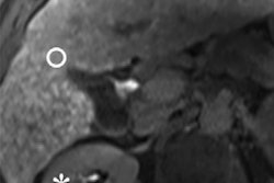
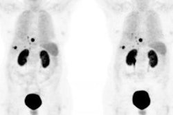
Researchers from the University of California, Los Angeles (UCLA) and the University of Pennsylvania are reporting success with a new PET tracer designed to image T-cells in the liver. The preclinical research was published in the October issue of the Journal of Nuclear Medicine.
Using a mouse model, the researchers observed how the F-18 FAC radiotracer can be used to image T-cells as they converge on the liver or during treatment with an immunosuppressive drug (JNM, October 2018, Vol. 59:10, pp. 1616-1623).
Physicians may look for T-cells to determine whether an organ is under attack and whether a biopsy is necessary. This can be useful in patients who have received an organ transplant, for example, and take immunosuppressant medication. Ultimately, the researchers hope that information gleaned from PET imaging with F-18 FAC will help avoid invasive and painful liver biopsies.
"The personalized treatment of patients suffering from immune attack on the liver will require precise and quantitative methods for measuring T-cells in the liver," said study co-author Peter Clark, PhD, from UCLA in a release from the Society of Nuclear Medicine and Molecular Imaging (SNMMI). "We envision that if this approach is translated into the clinic, it could lead to fewer liver biopsies and more precise treatment of patients with immune-related liver disease."


