University of Wisconsin researchers have used functional MRI (fMRI) for the first time to see the effects of antidepressant medication before and after treatment in patients with significant depression.
The study used fMRI of depressed patients and healthy subjects to examine changes over the course of antidepressant treatment in patterns of neural activity evoked by eliciting affective responses with affective visual stimuli.
Twelve patients with major depressive disorder and five healthy comparison subjects were scanned on three occasions, during which trials of alternating blocks of affective and neutral pictorial visual stimuli were presented, before treatment and after two weeks and eight weeks of treatment with venlafaxine (Effexor, Wyeth, Madison, NJ).
Baseline studies of both groups showed bilateral activation in the visual cortices, lateral prefrontal cortex, and the amygdala in response to the negative stimuli, the researchers wrote in the American Journal of Psychiatry, with patients showing greater activation in the visual cortex and less activation in the left lateral prefrontal cortex (January 2003, 160:1, pp. 64-75).
Symptoms were evaluated at each testing, and both groups completed self-report measures of mood. Statistical parametric mapping was used to examine the fMRI data with a focus on the group-by-time interactions.
The image acquisition protocol consisted of 10 scans, which included the following:
- a sagittal whole-brain T1-weighted spin-echo scan
- an axial three-dimensional spoiled gradient-recalled echo scan
- a three-dimensional time-of-flight scan
- a coronal three-dimensional spoiled gradient-recalled echo scan
- a coronal T1-weighted spin-echo scan
- a coronal gradient-echo dual-echo scan
- a coronal T2-weighted gradient-echo echo-planar scan
Scan 5, with one slice centered on the amygdala, provided the slice locations from which functional image data was acquired. The posterior edge of this slice was positioned 1 mm anterior to the location where the hippocampus could first be identified in the image data acquired in scan 4.
Image data were acquired on a 1.5-tesla EchoSpeed scanner (GE Medical Systems, Waukesha, WI) equipped with high-speed, whole-body gradients and a standard clinical whole-head transmit-receive quadrature birdcage head coil.
After two weeks of treatment with venlafaxine, the researchers observed significant increases in activation in the left insular cortex in patients versus comparison subjects. While the patients showed relatively less baseline activation in this region in response to negative stimuli than the comparison group did, after two weeks of venlafaxine treatment this difference was eliminated.
 |
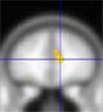
| These two cardinal views (coronal on the left; sagittal on the right) display a statistically significant interaction between group and time. The line graphs decompose the interaction, and reveal the increased anterior cingulate activation for treated patients, and a decrease in activation in this region among the controls, from baseline to eight-week assessment. Images courtesy of Dr. Richard Davidson and the University of Wisconsin. |
A similar change was found for the left anterior cingulate, where patients showed an increase in activation following treatment. Both regions have previously been implicated in the circuitry of emotion and its regulation.
That depressed patients had relatively less activation in these regions at baseline in response to negative stimuli is consistent with the theory that during an acute bout of depression, during an acute depressive bout, patients with major depressive disorder are hyporesponsive in the central circuitry associated with affective processing.
Patients with greater relative anterior cingulate activation at baseline in response to the negative versus neutral stimuli showed the most robust treatment response.
The patients showed a mean 70% reduction in Hamilton depression scale scores from the initial assessment to the eight-week assessment. This was the first study to show reliable MR signal changes in response to emotion activation in depressed patients with antidepressant medication. Although the study group was small, a number of predicted differences were found.
By Robert BruceAuntMinnie.com contributing writer
February 28, 2003
Related Reading
Functional MRI shows how brain lets you smell that smell, February 14, 2003
Virtue its own reward, fMRI confirms, August 22, 2002
New fMRI studies show learning and judging in action, November 15, 2001
Neuroscientists map physiology of feelings, September 20, 2000
Copyright © 2003 AuntMinnie.com