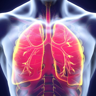
Diffusion-weighted MRI (DWI-MRI) offers comparable or even superior diagnostic performance to FDG-PET/CT for differentiating malignant and benign pulmonary nodules and masses, according to a meta-analysis published online November 27 in Radiology.
After analyzing some three dozen articles in the literature, researchers from the U.S. and Brazil found that DWI-MRI yielded a statistically significant improvement over FDG-PET/CT in sensitivity, diagnostic odds ratio, and area under the receiver operating characteristic curve for characterizing indeterminate lung nodules.
"This meta-analysis supports the inclusion of [DWI-MRI] as a radiation-free and less costly imaging modality for the primary noninvasive diagnostic workup of suspicious pulmonary lesions," wrote lead author Dr. Adriano Basso Dias, from Pavilhão Pereira Filho Hospital in Porto Alegre, Brazil, and colleagues.
FDG-PET/CT combines metabolic and morphologic information to characterize pulmonary nodules, but the modality can misclassify benign inflammatory nodules and can't always detect some well-differentiated pulmonary adenocarcinomas, according to the researchers. In addition, it's costly and exposes patients to radiation. DWI-MRI, however, can differentiate between normal and abnormal tissue for a variety of conditions by quantifying the flow of water molecules.
"The few studies that compared [DWI-MRI] and PET/CT for differentiation of malignant from benign pulmonary nodules demonstrated similar accuracies between the two modalities," the authors noted.
To their knowledge, there had been no meta-analyses on this topic. Therefore, the researchers sought to perform a systematic review and meta-analysis of the literature to compare the diagnostic performance of the two modalities. They searched the Pubmed, Embase, and Cochrane databases up to December 2017 and found 37 English-language articles eligible for their review.
The research studies included 23 papers involving FDG-PET/CT, eight assessing DWI-MRI, and six with both modalities. Of the research using FDG-PET/CT, the total sample size was 3,867 patients with 4,075 pulmonary lesions. In the studies that compared DWI-MRI and FDG-PET/CT, the total sample size was 651 patients with a total of 728 lesions (77% malignant, 23% benign).
To reduce interstudy heterogeneity, the researchers elected to focus primarily on the six research studies in which both DWI-MRI and FDG-PET/CT were performed on the entire patient population. They evaluated lesion-to-spinal cord signal-intensity ratio and apparent diffusion coefficient for DWI-MRI, and maximum standardized uptake values for PET/CT. Next, the researchers calculated pooled diagnostic performance statistics for the six studies.
| Pooled diagnostic performance for evaluating indeterminate pulmonary lesions | ||
| FDG-PET/CT | DWI-MRI | |
| Sensitivity | 78% | 83% |
| Specificity | 81% | 91% |
| Diagnostic odds ratio | 15 | 50 |
| Area under the curve | 0.86 | 0.93 |
The differences in sensitivity, diagnostic odds ratio, and area under the curve were all statistically significant (p = 0.018, 0.001, and 0.006, respectively), while the increase in specificity approached significance (p = 0.056).
"Despite the heterogeneity and limitations found in this study, these data suggest the need for larger-scale randomized controlled trials for the direct comparison of [DWI-MRI] and FDG-PET/CT in the setting of suspicious lung pathologic findings," the authors wrote.