Radiologic technologists require time and practice to fully develop their ability to acquire high-quality x-ray images on the first attempt, but chances to gain clinical experience are limited because the modality involves exposing participants to radiation, Yongsu Yoon, PhD, told AuntMinnie.com.
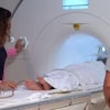
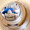
To increase the opportunities for practice, researchers from Kyushu University in Fukuoka developed a radiographic simulator that uses the Microsoft Kinect motion sensor to provide technologists with a safe and cost-effective way to improve their x-ray imaging prowess with real-time detection and image processing.
"Our proposed real-time radiographic simulator can display the simulated image in a way that is very close to actual radiography in real-time," Yoon said. "It is mainly focused on the education and training of beginner-level radiologic technologists in medical institutions, but it can also be helpful for radiologists to understand the relationship between 2D anatomic structure on an image and real-time shifts in patient positioning."
The researchers will describe the logistics of using the simulator and also the optimal settings for the device in this digital education exhibit.