Dr. W. Thomas Gregory, of Oregon Health and Science University, and colleagues used MRI to evaluate changes in the pelvic floor before and after a first pregnancy, comparing these changes between women who had a cesarean delivery and those who had a vaginal delivery.
Forty-two women had a baseline evaluation before pregnancy, six weeks after delivery, and six months after delivery. At all three visits, each woman had an interview with a questionnaire, a clinical pelvic exam, transperineal and endoanal 3D ultrasound, electromyography of the pelvic floor and anal sphincter muscles, and pelvic MRI using a 3-tesla magnet.
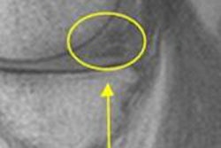
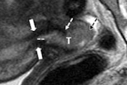
The researchers focused on the MRI findings of the women who had completed visits 1 and 2 (baseline evaluation and six weeks after delivery). They found significant differences in soft-tissue measurements of the pelvic floor, including statistically significant inferior position of the bladder neck six weeks after delivery in all women. The finding was more pronounced following vaginal delivery compared to cesarean delivery, the group noted.
This was the first study to image pelvic structures in the same woman before and after delivery, Gregory and colleagues believe. The results may help physicians and patients better predict the risks and benefits of cesarean delivery.