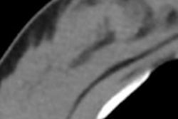
Dr. Reni Butler, from Yale School of Medicine, and colleagues reviewed 155 breast cancers diagnosed between October 2011 and January 2013 that were imaged with tomosynthesis in combination with 2D mammography. Seven breast radiologists evaluated the images, classifying them into five categories:
- Only seen on tomosynthesis
- Better seen on tomosynthesis
- Equally well seen on both modalities
- Better seen on mammography
- Only seen on mammography
The reading radiologists also recorded breast density, type of mammographic finding, clinical presentation, and cancer histology for each case.
Butler and colleagues found that women in the middle of the breast tissue density continuum had the highest percentage of cancers seen only on or better with tomosynthesis, with a rate of 67.7% for scattered fibroglandular tissue and 69.8%% for heterogeneously dense tissue. Tomosynthesis alone visualized only 5.9% of cancers in patients with extremely dense breasts and 0% in patients with fatty breast tissue.
Patients with fatty breasts and extremely dense breasts had the most cancers seen equally well on either modality, at 86.7% and 58.8%, respectively. As tomosynthesis becomes more widely used, it's crucial for radiologists to understand its relative benefit in different groups of patients, Butler's group concluded.