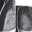
Digital breast tomosynthesis outperformed conventional mammography for detecting early cancers -- particularly those that present as architectural distortions, Dr. Elizabeth Rafferty and colleagues found.
The study included 172 cancers identified on imaging with both tomosynthesis and mammography between March 2011 and October 2012. Rafferty's group recorded each cancer's visibility (using a five-point scale), as well as its shape and margins as depicted by two-view tomosynthesis and mammography images.
Of the 172 cancers, 142 were invasive ductal carcinoma, 25 were invasive lobular carcinoma, and five were invasive mammary carcinoma. Tomosynthesis was more effective in visualizing invasive ductal carcinoma and invasive lobular carcinoma than mammography, with scores of 3.4 versus 2.6 and 3.2 versus 2.3, respectively. And tomosynthesis identified more cancers: 16% of all cancers were not identified by mammography, according to Rafferty's team, while only 3% were not identified by tomosynthesis.
In addition, of the cancers that appeared as architectural distortions on tomosynthesis, 50% were hidden on conventional mammography and 20% were characterized as asymmetry or focal asymmetry on conventional mammography. Based on the results, Rafferty and colleagues concluded that tomosynthesis may help clinicians better assess cancers at initial imaging.
