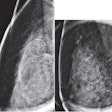
Though FFDM has been shown to be more accurate than film-screen mammography in women with dense breasts, identifying amorphous calcifications is often difficult with either method, said presenter Anabel Scaranelo, MD, PhD. As a result, the researchers sought to evaluate the use of CAD with FFDM in this application.
The study team retrospectively applied CAD to FFDM images of 122 women with amorphous calcifications; the group found that CAD identified all 36 amorphous calcifications that turned out to be malignant. In addition, CAD yielded 85% sensitivity in detecting lesions deemed to be high risk and 80% sensitivity in benign amorphous calcifications.
The use of CAD directly with FFDM was much more effective than when the technology is applied to digitized films, Scaranelo said.
"This is very important because of the issue of reliability and reproducibility of the method," she told AuntMinnie.com.
In other results, Scaranelo will describe how CAD markings resulted in a 29% likelihood of malignancy, a higher figure than found in previous studies.
"When we correlated with patient risk factors, we found amorphous calcifications [to be] 1.76 times more likely to be malignant or show 'high-risk' lesions in survival patients or women with prior breast biopsy showing [atypical ductal hyperplasia] in the contralateral breast," she said. "This emphasizes the need for early detection of precancerous lesions, particularly among amorphous calcifications."


