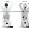
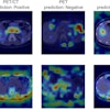
Wendie Berg, MD, PhD, and colleagues from Johns Hopkins at Green Spring Station in Lutherville, MD, will explain findings from their study that compared PEM's performance to whole-body PET and PET/CT for additional cancer detection, presurgical planning, and axillary staging.
Berg's team used data from a previous study conducted between September 2006 and November 2008, during which 388 women with newly diagnosed breast cancer who had been offered breast-conserving surgery underwent both bilateral contrast-enhanced MRI and F-18 FDG PEM. Of this 388, 179 also had whole-body PET (71 of 179) or whole-body PET/CT (108 of 179). Median index tumor size was 1.4 cm.
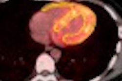
Of the women who had PET/CT and PEM, all had residual tumors at surgery. PEM found 103 tumors (95.4%) and PET/CT found 94 (87%). In addition, 23 of these women had additional ipsilateral cancers: PEM found 13 of these and PET/CT found three.
Of the women who had PET and PEM, 68 (96%) had residual tumors at surgery. PEM caught 63 (93%) of these 68 tumors, while PET caught 38 (56%). In addition, 15 of these women had additional cancers on the opposite breast: PEM found seven of these (47%) and PET/CT found one (6.7%).


