Seeking to evaluate the diagnostic value of ultrasound in this patient population, the team performed a six-month prospective study to evaluate all children presenting at the University of Jena's pediatric emergency department with clinical suspicion of pneumonia, said presenter Dr. Martin Stenzel.
The study included 50 children who were examined with both ultrasound and x-ray of the thorax. Two experienced pediatric radiologists compared the results of both methods.
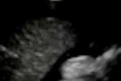
Ultrasound was able to detect more pulmonary consolidations, which are found in bacterial pneumonia, than x-ray. In addition, ultrasound revealed additional information in 18 hemithoraces.
On the other hand, ultrasound did not detect centrally located consolidations that were found on x-ray in 19 lungs, Stenzel said.
Stenzel believes the study's conclusions will be interesting to both pediatricians and pediatric radiologists. The results show that a well-trained examiner can delineate pulmonary consolidations and tissue hyperemia on sonography, and also assess for pleural effusion, he said.
"Children with suspected pneumonia can be assessed by a meticulous ultrasonography of the thorax first, which often yields the diagnosis already," Stenzel said. "In cases of obvious disparity, the chest x-ray examination is still valuable as a second-line method."
Multi-institutional prospective studies are now needed to validate this approach, especially considering the variability in skills of sonographic examiners, Stenzel noted.