The tool was internally developed and is tightly integrated with the department's PACS and its speech recognition system. In this scientific presentation, Dr. Paul Chang, professor of radiology and vice chair of radiology informatics at the university, will describe how the tool works and what it has accomplished.
Serial measurement of reference lesions is important to track their growth or their shrinkage as they respond to treatment. But the process of identifying, comparing, measuring, and documenting these changes can be inefficient for radiologists and is prone to error.
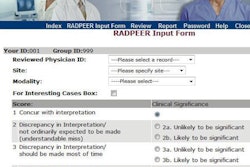
The tool automatically displays the image that best demonstrates the lesion on the prior exam in conjunction with the corresponding image from the current study. It also displays a lesion report table that is automatically populated with the lesion measurements after the user measures the lesion on the current study. Measurements are automatically entered into the radiology report.
Chang and colleagues used time-motion analysis to evaluate the efficiency of abdominal radiologists interpreting exams of cancer patients with the tool and without it. The radiology reports they created were also analyzed with respect to the number of errors made. The study showed very positive results, according to Chang. Learn what they were at this session.