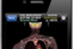
Radiologists use image annotations on PACS as part of their routine diagnostic interpretation process, and these annotations contain important visual information that can significantly augment radiologists' text reports. For various reasons, however, referring physicians can frequently only access the text-based report, said Sharmili Roy, who will present the collaborative effort from the National University of Singapore and Weill Cornell Medical College.
To enhance radiology reporting, the team sought to leverage the AIM standard, which creates and stores annotations in a manner that supports automatic searching and utilization of annotation and image content. Building upon AIM, the team developed Visual Interpretation With Three-Dimensional Annotations (VITA) to help in visually communicating these important clinical findings, Roy said.
VITA automatically extracts image-based annotations and produces an animated 3D summary in the form of a rotating 3D volume that clearly shows the annotations, Roy said. It can be accessed on PACS by the ordering provider or later by the radiologist.
"Our long-term vision is that this 'visually enhanced report' will become a common feature in radiological reporting," Roy said.