Dr. Gregor Dovjak, from the department of biomedical imaging and image-guided therapy at the Medical University of Vienna, is scheduled to outline the benefits of DTI for assessing cerebellar white-matter connectivity, which plays a significant role in a person's cognitive and motor skills.
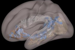
In this study, Dovjak and colleagues retrospectively looked at 14 fetal MRI scans from a 1.5- or 3-tesla system to gauge DTI's ability to visualize the superior, middle, and inferior cerebellar peduncle and the transverse pontine fibers. With the help of tractography software (IntelliSpace, Philips Healthcare), they created a visibility score based on the fraction of visible tracts divided by the amount of potentially visible tracts.
They were able to adequately discern the superior and middle cerebellar peduncle in approximately 70% of cases, the inferior cerebellar peduncle in slightly more than half the cases, and the transverse pontine fibers in all but one fetus.
The researchers concluded that prenatal tractography of cerebellar white-matter tracts showed "excellent correlation with the respective anatomy," adding that fetal DTI might help characterize any abnormalities in a fetus' forebrain before birth.