TSC causes nonmalignant tumors to grow in the brain, heart, lungs, kidneys, eyes, and skin. The condition can prompt seizures and behavioral issues and slow fetal development.
In a study from Hospital Universitari Vall d'Hebron in Barcelona, researchers evaluated 12 fetuses between the 22nd and 36th week of gestation with cardiac rhabdomyomas (a tumor of the heart) by having them undergo cerebral MRI.
The MRI protocol included a body phased-array coil, and images were obtained using a variety of MRI sequences, with women imaged in the supine position without sedation.
In 10 cases, fetal MRI demonstrated typical characteristics of tuberous sclerosis complex, which were confirmed by postnatal MRI in pregnancies that continued gestation or via histology in those that were interrupted.
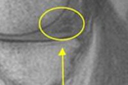
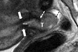
"The benefit of MRI is that it can discover very early brain lesions of tuberous sclerosis complex -- mainly cortical tubers and subependymal nodules -- usually not apparent on a detailed sonographic examination," said lead author Dr. Élida Vázquez, chief of pediatric neurology. "The presence or absence of cerebral lesions in suspected fetal tuberous sclerosis complex, as well as the number of these lesions, crucially influences the prognosis and gestation management."
Fetal MRI is beneficial for detecting cerebral lesions in tuberous sclerosis complex and should be considered as part of a diagnostic workup in suspected TSC cases, the researchers concluded.