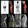
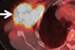
Given these results, study presenter Dr. Kate Hanneman, an assistant professor of radiology at the University of Toronto, said that simultaneous PET/MRI could potentially reduce the number of exams for cardiac patients. Hanneman performed the study during a fellowship at Stanford University.
This particular study investigated PET/MRI with time-of-flight (TOF) imaging of patients with cardiac sarcoidosis, an inflammatory disease that can affect heart function and has been increasingly cited as a cause of heart failure and arrhythmias.
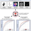
Hanneman and colleagues prospectively recruited six patients over three months to evaluate the subjects for suspected cardiac sarcoidosis using standard clinical evaluation and simultaneous PET/MRI. Five healthy volunteers also were scanned as a control group.
The patients first underwent standard cardiac PET/CT (Discovery 600 or 690, GE Healthcare) after administration of FDG. This was followed by simultaneous PET/MRI with a 3-tesla scanner and TOF imaging (Signa, GE Healthcare). The total scan time for PET/MRI was significantly longer than for PET/CT.
PET images from the PET/CT scans were positive for cardiac sarcoidosis in 50% of patients, while PET from PET/MRI exams were positive for cardiac sarcoidosis in all of the patients, the researchers found.
They also found a significant difference in effective radiation dose between the two hybrid modalities, with, predictably, much less radiation from the PET/MRI scans.
Based on the results, cardiac PET/MRI with TOF is feasible for "achieving diagnostic image quality, with the added benefit of radiation dose reduction in comparison to PET/CT," the group concluded.
