"With regard to patient comfort related to scan duration and a markedly reduced radiation exposure, FAST PET/MRI may serve as a powerful alternative to PET/CT for diagnostic workup of patients with lymphomas," said lead author Dr. Johannes Grueneisen from University Hospital Essen.
In this study, more than 40 consecutive lymphoma patients were prospectively enrolled for a clinically indicated PET/CT exam and a follow-up PET/MRI scan. The FAST protocol consists of a whole-body diffusion-weighted echo-planar imaging (EPI) sequence, a half-Fourier acquisition single-shot turbo spin-echo (HASTE) sequence, and a postcontrast 3D volumetric interpolated breath-hold examination (VIBE) sequence.
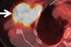
The researchers then calculated standardized uptake values (SUVs) for all FDG-avid lesions identified on PET/CT and PET/MRI. Lymphoma was found in 24 patients, with both hybrid modalities correctly identifying all malignant lesions in each patient. In addition, all PET-positive lesions visible on PET/CT were also seen on PET/MRI.
In the future, the researchers hope to evaluate the diagnostic capability of integrated PET/MRI for monitoring radiotherapy and/or chemotherapy for lymphoma patients.
"Lymphoma patients might particularly benefit from this new imaging modality, considering the percentage of a younger patient population and the need for repetitive examinations, potentially increasing the risk of radiation-associated second malignancies," Grueneisen told AuntMinnie.com.