The study from Karolinska University Hospital and the Karolinska Institute also found that MRI without contrast is insufficient in this clinical application.
"We found that CT provides the best possible means for staging of lymph node stations, both N and M staging in our specific patients," lead researcher Dr. Michael Torkzad, PhD, told AuntMinnie.com.
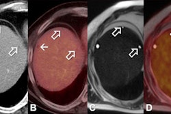
Torkzad and colleagues evaluated 35 patients with histologically confirmed squamous cell carcinoma of the anorectum and anal verge, who also received MRI, contrast-enhanced CT prior to FDG-PET/CT, and biopsies.
CT obtained the best set of criteria to assess lymph node staging, based on factors such as the short-axis diameter of the lymph node, a clear sign of necrosis, and the density of the node. Using the criteria, sensitivity and specificity with CT were 100%.
The results provide "criteria to stage lymph node station in these patients on CT," Torkzad said. "Also, PET/CT is not necessary for staging of these patients, if you are an expert reader of MRI or CT."
The study's findings should reinforce that lymph node staging in anal cancer is different from lymph node staging in other gastrointestinal cancers, he added.
Torkzad and colleagues plan to see if these results can be confirmed with other types of squamous cell cancer, such as head and neck cancer and cervical cancer.