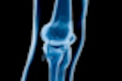
Lead author and presenter Dr. Thomas Magee, managing partner at Neuroskeletal Imaging in Merritt Island, FL, noted that isotropic MRI of the knee has been limited due to long acquisition times, issues with specific absorption rates (SARs), and z-axis constraints. With the help of phased-array coils, fast gradients, high-field 3-tesla MRI, and parallel imaging, high-resolution isotropic images can be acquired in less than six minutes.
"MRI now has the capability to produce high-quality isotropic MR imaging, as has been true of CT for years," Magee said. "These high-quality images are equivalent and in many ways superior to traditionally acquired 2D images. This allows the MR reader to view pathology in any given plane."
Two experienced musculoskeletal radiologists prospectively interpreted 3-tesla MR images of the knee in 50 consecutive patients, who also had XETA images with 3D reconstruction. In addition, 39 of the 50 patients underwent subsequent arthroscopy. Results then were compared with MR interpretations.
The analysis showed that 36 (72%) of the 50 patients had meniscal tears on prospective conventional MRI and XETA MRI. Of these meniscal tears, 21 were nondisplaced horizontal tears, six were bucket handle tears, five were radial tears, and four were displaced flap tears.
Meniscal tears were diagnosed with equal accuracy on conventional MRI exams as with 3D XETA imaging, the researchers also concluded. In addition, the exact location and degree of displacement of meniscal tears were more accurately diagnosed on isotropic and 3D XETA acquisitions than with conventional MRI.
Arthroscopic findings correlated with MRI findings in 34 (94%) of the 36 patients.
Magee and colleagues plan to follow up the study by performing 3D isotropic imaging routinely for all patients. "It is our belief that this provides accurate, high-resolution images in a shorter time frame, which allows for better patient tolerance of the procedure," he said. "Also, the MR reader can view pathology in any plane, allowing for equal and probably more accurate diagnoses as compared to traditional 2D imaging."