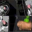
Few radiology departments currently use interventional phantoms, but there is tremendous opportunity now to create low-cost, noncommercial models using 3D printing technologies, said presenter Dr. Ramin Javan. 3D printing, or rapid prototyping, is a technique for building solid objects in a layer-by-layer fashion.
Patents on fused deposition modeling ran out a few years ago, paving the way for the emergence of ultralow-cost desktop 3D printers. Laser sintering technologies also will soon come off patent protection, enabling an even greater increase in 3D printing applications due to higher accuracy, Javan said.
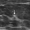
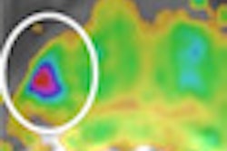
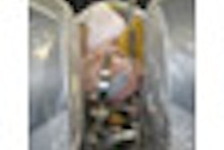
As gatekeepers of medical imaging -- especially cross-sectional techniques -- radiologists have a real opportunity to apply this technology in all aspects of medicine, Javan said. Using 3D printing and molding techniques with a variety of materials, he was able to create a prototype dual-modality interventional phantom of the neck.
"This model can be used for practicing neck interventional procedures multiple times without the anxiety of potentially harming any patients in the process and gaining confidence in one's techniques for performing a certain procedure," he told AuntMinnie.com. "The techniques and materials used and introduced also may serve as the backbone of other potential applications for investigators, such as developing custom intraoperative drill guides or customized prosthetics."
Javan said he is also developing interventional phantoms of the sinonasal system, skull base, face, abdominal vasculature, and liver. In addition, Javan has founded the company, 4D-Inceptions.


