However, diffusion-weighted MRI (DWI-MRI) can be a valuable adjunct to T2-weighted images and comparable to contrast-enhanced T1-weighted images in detecting neuroendocrine tumors of the pancreas.
Researchers led by Dr. Carsten Rist, an associate professor of radiology and the section chief of PET/CT, analyzed 18 patients with proven pancreatic neuroendocrine tumors who underwent DWI-MRI and contrast-enhanced PET/CT within six weeks. Two radiologists then compared the results of MR imaging techniques and PET/CT.
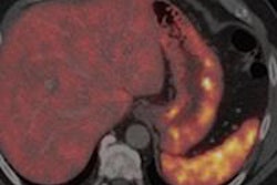
With gallium-68 DOTATATE PET/CT, both readers were able to detect all 23 pancreatic neuroendocrine tumors, for a detection rate of 100%. By comparison, one reader found only eight (35%) neuroendocrine tumors and the other reader found nine (39%) using T2-weighted MRI.
The detection rate improved significantly by combining T2-weighted MRI with DWI or contrast-enhanced T1-weighted imaging. One reader was then able to detect 14 neuroendocrine tumors (61%), while the other detected 15 tumors (65%).
Based on the results, Rist and colleagues concluded that DOTATATE-PET/CT should be the method of choice for diagnostic imaging of pancreatic neuroendocrine tumors, and that DWI is advantageous, especially if PET/CT is not available or contrast-enhanced imaging is contraindicated.