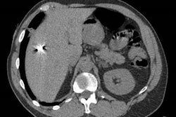
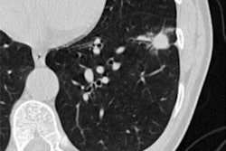
Stefan Sawall, PhD, from the German Cancer Research Center in Heidelberg will discuss findings on PCCT as a newcomer in breast screening. Sawall and colleagues used breast and thorax phantoms to represent realistic patient positioning. PCCT dose levels varied between 7 mGy and 25 mGy (CT dose index volume [CTDIvol]) at 120 kV in ultrahigh resolution mode. Meanwhile, images for dedicated CT were obtained using a standard protocol with a dose of 7 mGy (CTDIvol) at 49 kV.
The researchers wanted to compare the two methods by evaluating contrast-to-noise ratio of masses, the noise power spectrum, and the structural visibility of fibers and calcifications. They found that the contrast-to-noise ratio between masses and background was 1.3 on average when only using the breast phantom. Including the thorax phantom though, these differences varied between 0.9 to 2.3 mGy.
The researchers also found that the noise power spectrum's peak frequency in PCCT was 0.45 mm-1 compared with 0.40 mm-1 in dedicated breast CT. Additionally, calcifications down to a size of 0.29 mm and fibers down to a size of 0.23 mm could be identified in both systems' respective images.
Sawall et al suggested that PCCT could become an alternative to dedicated breast CT by providing additional information, such as identification of sentinel lymph nodes. They called for more studies on dosage delivered to other organs.
Find out more in Sawall's presentation.


