The results suggest that photon-counting CT is a viable addition to the CT imaging toolkit, according to Dr. Martine Remy-Jardin, PhD, of the University Centre of Lille in France and colleagues.
"PCCT acquisitions provided similar morphologic image quality and superior perfusion imaging at lower radiation dose [compared to dual-source CT]," the group noted.
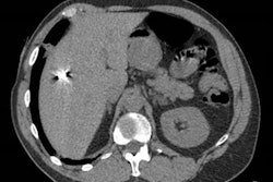
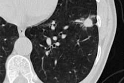
Remy-Jardin's team compared image quality of photon-counting CT and dual-source CT for chest CT angiography examinations in a study cohort of 142 patients. Half were scanned with a photon-counting CT device and half with a dual-source CT system.
The mean dose length product of photon-counting CT was significantly lower than dual-source CT, at 172.6 mGy.cm compared to 339.4 mGy.cm (p < 0.0001) -- a 52% reduction. The researchers also found that photon-counting CT exam time was shorter compared to dual-source CT, at 0.93 seconds compared to four seconds.
The group found no significant difference between the two techniques when it came to mean attenuation value in the central pulmonary arteries; background noise; or mean level of attenuation on perfusion images.
"Photon-counting CT has the potential to improve perfusion imaging while maintaining morphologic image quality with a radiation dose reduced by 50%," the investigators concluded.