Clinical trials have shown that clinical benefit for the patient with coronary artery disease is achieved by only intervening on lesions that are hemodynamically relevant, i.e., those that reduce myocardial blood flow. Intervention on functionally irrelevant lesions has the potential to cause more harm than good.
"The CT techniques discussed in this session are exciting, because they enable us to noninvasively identify stenoses that indeed cause lesion-specific ischemia," presenter Dr. U. Joseph Schoepf told AuntMinnie.com.
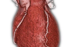
Those techniques include CT myocardial perfusion imaging, CT-derived fractional flow reserve, and their precursor methods, such as transluminal attenuation gradient measurements.
"With all these techniques, the goal is to analyze coronary artery anatomy and function with the same modality, obviating the need of subjecting the patient to multiple testing," Schoepf said. "Initial results are highly promising."
Studies have shown that by applying those methods, the specificity and positive predictive value of coronary CT angiography can be improved, thus reducing lesion overcall and unnecessary invasive workups, according to Schoepf. Exciting new data to be presented indicate that functional coronary CT imaging is achieving a coveted goal among medical imaging tests -- namely, a measurable improvement in clinical outcomes and quality of life, along with a reduction in healthcare costs.
"These new tests are prime examples of the volume-to-value proposition and will play a pre-eminent role in the future of our field," he said.