Searching for the lowest possible dose is ideally done by scanning the same body region multiple times using different dose levels, but multiple scans raise ethical challenges, leading most investigators to turn to phantoms, according to Dr. Stefan Wirth from Ludwig Maximilian University of Munich.
However, using cadavers is a better choice because they make the best possible phantoms and enable multiple scans to be performed without ethical questions.
"The purpose of this study was to explore the lower end of possible dose rate reduction, while keeping image quality at least stable/diagnostic with the help of fully iterative algorithm modeling at different dose levels in contrast-enhanced MDCT of the chest," Wirth wrote in an email to AuntMinnie.com.
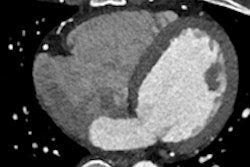
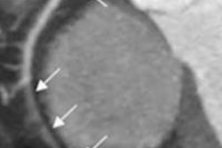
In the study of 11 cadavers, model-based iterative reconstruction (MBIR, GE Healthcare) reduced dose by 82% compared with an earlier reconstruction algorithm, adaptive statistical iterative reconstruction (ASIR, GE), leading to submillisievert chest scans for all cadavers without loss of image quality.