Earlier research showed that the cumulative frequency of low-attenuating lung regions follows a power law characterized by exponent D, yet another study appeared to contradict those results, saying that it was differences in the attenuation threshold that explained the discrepancy, explained Dr. Mizuho Nishio from the Kobe Graduate School of Medicine.
In their study, Nishio and colleagues aimed to validate the power rule for low-attenuation 3D clusters in the lung and, by doing so, demonstrate a reliable method of COPD assessment.
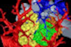
Twenty-five patients underwent CT on a 320-detector-row scanner with 1-mm-thick reconstructions. Percentages of lung regions with low-attenuation 3D clusters were computed and the frequency distribution evaluated by linear regression. The low-attenuation percentage and exponent D were calculated at 20 different attenuation thresholds.
Based on linear regression, Pearson's correlation coefficients ranged from -0.99 to -0.83, corroborating the validity of the power law (D) for the size distribution of 3D low-attenuation clusters.
"D shows significant correlation with results of pulmonary function testing when the thresholds for low-attenuation lung regions are properly chosen," Nishio told AuntMinnie.com. "This result shows that using D pulmonary emphysema can be quantified accurately."