Radiological estimates of tumor blood flow are important both for diagnosing disease and choosing therapies, Dr. Davide Ippolito, from the University of Milano-Bicocca, told AuntMinnie.com.
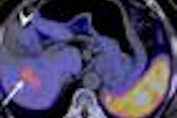
In the liver, the source of blood supply differs between tumor and normal tissue. The two blood supply sources -- the portal vein and the hepatic artery -- are altered in many diseases, he said. Thus, perfusion parameters as measured with functional CT can be used as physiologic markers that are related to the vascular changes induced by tumor neoangiogenesis.
Angiogenesis can be used as a prognostic indicator to predict the outcome of the disease and the best treatment. "Tumor growth cannot be sustained without an adequate blood supply, and drugs targeted at tumor vasculature are an attractive therapeutic strategy, which has already yielded real patient benefits," Ippolito noted.
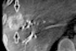
The researchers analyzed 56 biopsy-proven HCC lesions and 41 HCC lesions treated with transcatheter arterial chemoembolization (TACE) or radiofrequency ablation. The CT study acquired eight dynamic slices per scan at a fixed table position during injection of nonionic contrast.
The results show that CT perfusion can noninvasively quantify tumor blood supply related to the formation of new arterial structures, the group will report. Perfusion CT is a relatively simple technique that could be integrated into the current CT protocols, providing an in vivo marker of tumor-related angiogenesis.