"Currently, invasive right-heart catheterization is required to confirm and often also to exclude a diagnosis of pulmonary hypertension," said presenter Dr. Fabian Rengier, a research fellow at the University of Heidelberg who won a trainee award for the study.
Pulmonary artery dimensions are known to be greater in patients with pulmonary hypertension. However, previous studies focusing on 2D axial measurements yielded only moderate sensitivity and specificity for predicting pulmonary hypertension, according to Rengier.
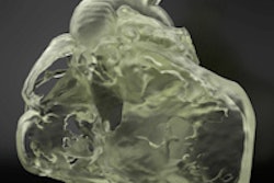
The investigators acquired MR angiography images on a 1.5-tesla scanner in patients with pulmonary hypertension and in normal volunteers. Using analytical software developed in house, they segmented the main, left, and right pulmonary arteries automatically after placing seed points. Volumes for the arteries were computed and corrected for body surface area, and the diameter of the main pulmonary artery was manually measured for comparison purposes.
3D pulmonary artery volumes may be more accurate than 2D diameter measurements for predicting and assessing pulmonary hypertension, they concluded.
Learn more by stopping by this Wednesday session.