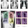
The use of chest radiographs to detect tuberculosis (TB) in areas with a high burden of TB is limited by the low number of skilled readers available. As a result, the researchers sought to develop software that automatically analyzes chest x-rays, detects abnormalities, and indicates the likelihood that these abnormal signs are consistent with TB and with active TB, said presenter Laurens Hogeweg of University Medical Center Utrecht in the Netherlands.
In testing, the CAD software produced an area under the receiver operator characteristics (ROC) curve of 0.81. While the prototype is being improved, the research team is already planning a number of larger observer and clinical studies to further evaluate the system's performance, Hogeweg said.
In the future, the software will be bundled with clinical x-ray machines used in the field in several countries in southern Africa. It can then play a role in facilitating large-scale tuberculosis screening programs, reducing the number of images that have to be read, he said.
"Another option is to use the software in daily clinical practice," Hogeweg told AuntMinnie.com. "The opinion of the software could be presented to a clinical officer, allowing a diagnosis of TB on the spot, and a TB suspect could immediately be put on treatment."