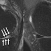
Adrenal vascular imaging is a relatively unexplored imaging area, mainly due to cross-sectional spatial resolution limitations when attempting to display such small-caliber vessels, said presenter Supriya Gupta, MD, of Massachusetts General Hospital (MGH) in Boston.
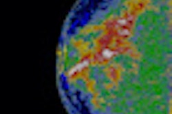
In this exhibit, researchers will discuss how existing MDCT protocols and various algorithms in 3D postprocessing techniques for adrenal imaging can be employed for visualizing adrenal vascular details, Gupta said.
While some MDCT imaging limitations remain due to the thin caliber of the adrenal vessels, their complex course, and frequent superimposition by surrounding structures, the authors will propose ways of countering these challenges. In addition, the group will highlight emerging and future applications of 3D postprocessing for mapping the adrenal vasculature, Gupta said.


