By Erik L. Ridley, AuntMinnie.com staff writer
    November 15, 2010

    The ever-increasing role that advanced visualization technologies play in today's radiology practice will be fully evident at this year's RSNA meeting in Chicago. Those seeking to keep up on the latest developments in 3D and computer-aided detection (CAD) will find a plethora of educational opportunities to choose from at McCormick Place.

    3D will be well-represented in scientific sessions, with highlights including new automatic segmentation techniques and presentations on how 3D quantitative analysis can reliably measure coronary plaque. A number of education exhibits also will feature important applications for 3D.

    In CAD, a broad range of scientific papers will be presented, covering the technology's use in a variety of lung applications, the prostate, the heart, the liver, and, of course, the breast. In virtual colonoscopy CAD, researchers are examining ways of improving the technology's utility and efficiency.

    And interest continues to surge around the use of smartphones and Apple's iPad tablet computer for viewing medical images.

    Refresher courses and other sessions

    Of course, RSNA educational opportunities don't just end with scientific sessions and education exhibits. Advanced visualization topics will be covered in a number of refresher courses and other sessions as well.

    In 3D, a talk on advanced imaging techniques will be the final presentation in a "Practical Informatics for the Practicing Radiologist" refresher course on Monday, November 29 (10:30 a.m.-12:00 p.m., II22C, Room S501AB).

    Another Monday course will focus on 3D interactive visualization of DICOM images for radiology applications (2:30 p.m.-4:00 p.m., IA22, Room S401CD).

    On Tuesday, November 30, an image processing and 3D imaging refresher course will feature talks on organ and tumor segmentation, 3D for education, and enterprise 3D tools to enhance your practice (8:30 a.m.-10:00 a.m.; RC330A, B, and C; Room S403A).

    Those sticking around until Friday, December 3, will be interested in an advanced visualization refresher course offered in association with the Society for Imaging Informatics in Medicine (SIIM). Presentations will cover 3D and advanced processing, how to select a 3D system for your environment, and ensuring the success of your 3D laboratory (8:30 a.m.-10:00 a.m.; RC826A, B, and C; Room S403B).

    A number of hands-on 3D workshops will also be held throughout the week.

    As for CAD, a minicourse on Sunday, November 28, will include talks covering a short history of CAD, engineering aspects of CAD, and the radiologist's perspective on CAD (2:00 p.m.-3:30 p.m.; RC123A, B, and C; Room S504CD). On Monday, November 29, another minicourse on CAD will address evaluation issues, including talks on clinical and technical evaluation issues; the limitations of CAD clinical studies; and the U.S. Food and Drug Administration's perspective (8:30 a.m.-10:00 a.m.; RC223A, B, and C; Room S504CD).

    Bright and early on Tuesday, November 30, a controversy session will tackle CAD for breast, lung, and colon cancer quantitative image analysis for clinical practice (7:15 a.m.-8:15 a.m., SC30, Room E351).

    Breast CAD enthusiasts won't want to miss a Tuesday refresher course, which will include talks on breast computer-aided detection, computer-aided diagnosis and beyond, and clinical aspects of computer-aided detection and computer-aided diagnosis (8:30 a.m.-10:00 a.m.; RC321A, B, and C; Room S503AB).

    Another CAD minicourse is on tap for Tuesday, featuring CAD applications in colon imaging and beyond (4:30 p.m.-6:00 p.m.; RC423A, B, and C; Room S504CD). Also on Tuesday, an additional refresher course will examine CAD for the rest of the body (4:30 p.m.-6:00 p.m.; RC430A, B, and C; Room E263).

    Other CAD-related refresher courses include a Thursday, December 2, CAD with tomosynthesis session, featuring talks on CAD with breast tomosynthesis, computerized detection of lung nodules on chest tomosynthesis, and optimizing the reading of digital breast tomosynthesis examinations (4:30 p.m.-6:00 p.m.; RC725A, B, and C; Room S404AB). A mammographic CAD presentation also will be given during a Friday, December 3, course on advanced breast imaging/technology (8:30 a.m.-10:00 a.m.; RC815A, B, and C; Room N228).
    Scientific and Educational Presentations
    Inexperienced readers benefit most from CAD in CCTA
    Sunday, November 28 | 10:55 a.m.-11:05 a.m. | SSA19-02 | Room S403B
    Researchers from the Medical University of South Carolina in Charleston will discuss how reader experience affects the benefits of computer-aided detection (CAD) in coronary CT angiography (CCTA).
    ASIR may bolster CAD performance in lung nodules
    Sunday, November 28 | 11:05 a.m.-11:15 a.m. | SSA19-03 | Room S403B
    In this scientific session, a team from Osaka University Medical School in Japan will present data detailing the benefits of adaptive statistical iterative reconstruction (ASIR) technology for computer-aided detection (CAD) of lung nodules.
    CAD offers potential in detecting tuberculosis
    Sunday, November 28 | 11:15 a.m.-11:25 a.m. | SSA19-04 | Room S403B
    A Dutch research group will share promising initial results from using prototype computer-aided detection (CAD) software to identify tuberculosis on chest x-ray images in this Sunday morning session.
    CAD may help detect pulmonary embolism
    Sunday, November 28 | 11:25 a.m.-11:35 a.m. | SSA19-05 | Room S403B
    A research team from the University of Michigan in Ann Arbor will share developments with its computer-aided detection (CAD) algorithm for detecting pulmonary embolism on CT pulmonary angiography.
    Automated 3D method may replace manual renal cortex segmentation
    Sunday, November 28 | 11:35 a.m.-11:45 a.m. | SSA09-06 | Room E351
    In this Sunday presentation, a research team from the U.S. National Institutes of Health (NIH) will share its success with an automated 3D renal cortex segmentation technique for abdominal CT images.
    Algorithm can automatically detect abnormal lung volumes
    Sunday, November 28 | 11:45 a.m.-11:55 a.m. | SSA19-07 | Room S403B
    In this paper presentation, researchers will provide details on a computerized method for identifying abnormal lung volumes from interstitial lung disease.
    CAD shows promise for evaluating prostate cancer aggressiveness
    Sunday, November 28 | 11:55 a.m.-12:05 p.m. | SSA10-08 | Room E353B
    Researchers from the University of Chicago will describe the potential for their prototype computer-aided detection (CAD) software in prostate MRI in this Sunday scientific session.
    Ultrasound CAD aids in differential diagnosis of focal liver lesions
    Sunday, November 28 | 12:05 p.m.-12:15 p.m. | SSA21-09 | Room S504AB
    In this scientific session, a team from Kumamoto University in Japan will present the benefits of computer-aided detection (CAD) in contrast-enhanced ultrasound of focal liver lesions.
    Evaluating the iPad's role in radiology
    Monday, November 29 | 10:50 a.m.-11:00 a.m. | SSC08-03 | Room S402AB
    In this scientific session, researchers from the University of Maryland in Baltimore will take a close look at the device generating significant buzz in the radiology community: Apple's iPad.
    iPhone holds its own against PACS workstation in telestroke cases
    Monday, November 29 | 11:10 a.m.-11:20 p.m. | SSC07-05 | Room S102D
    A team from Massachusetts General Hospital in Boston matched up an iPhone app and a PACS workstation for providing preliminary diagnosis on stroke evaluation cases, in this study to be presented Monday morning.
    Smartphones may support mobile teleradiology
    Monday, November 29 | 11:20 a.m.-11:30 a.m. | SSC08-06 | Room S402AB
    An Italian team will present promising results from using smartphones for mobile reading of CT and MR exams.
    Research weighs iPad clinical utility
    Monday, November 29 | 11:30 a.m.-11:40 a.m. | SSC08-07 | Room S402AB
    In this scientific paper presentation, a study team from the University of Maryland in Baltimore will describe how the iPad fared when used to evaluate a screening chest x-ray for tuberculosis.
    MPR CT images can demonstrate glenoid bone loss
    Monday, November 29 | 11:40 a.m.-11:50 a.m. | SSC10-08 | Room E451B
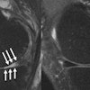
    In this paper presentation, Italian researchers will show how curved multiplanar reformatting (MPR) of CT images can quantify glenoid bone loss following shoulder dislocation.
    iPhone provides high image quality, but low speed
    Monday, November 29 | 11:50 a.m.-12:00 p.m. | SSC08-09 | Room S402AB
    A research team from Massachusetts General Hospital in Boston found both advantages and disadvantages to using an iPhone for preliminary diagnosis of chest CT pulmonary embolism studies.
    Chest x-ray CAD scheme suppresses ribs to improve results
    Monday, November 29 | 3:00 p.m.-3:10 p.m. | SSE06-01 | Room S404CD
    Researchers from the University of Chicago were able to improve the sensitivity of computer-aided detection (CAD) in detecting lung nodules -- presenter Sheng Chen will share their results in this Monday afternoon session.
    Bone suppression bolsters detection of pneumonia
    Monday, November 29 | 3:10 p.m.-3:20 p.m. | SSE06-02 | Room S404CD
    In another paper presentation from the University of Chicago, researchers will highlight the value of bone suppression imaging for improving detection of pneumonia on chest x-rays.
    Mammo CAD enhances interobserver agreement
    Monday, November 29 | 3:10 p.m.-3:20 p.m. | SSE21-02 | Room S403A
    In this scientific session, a research team from the U.S. Food and Drug Administration will discuss how adding ultrasound and computer-aided detection (CAD) to mammograms affects reader variability.
    3D quantitative analysis can measure coronary plaque
    Monday, November 29 | 3:30 p.m.-3:40 p.m. | SSE04-04 | Room S504AB
    In this Monday afternoon presentation, researchers will highlight the advantages of automated 3D quantitative coronary plaque analysis.
    New CAD approach reduces false-positives for HCC
    Tuesday, November 30 | 10:30 a.m.-10:40 a.m. | SSG13-01 | Room S403A
    A University of Chicago team will provide an update on its development of computer-aided detection (CAD) software for addressing hepatocellular carcinoma (HCC), according to this paper to be presented on Tuesday morning.
    Interactive breast ultrasound CAD improves lesion characterization
    Tuesday, November 30 | 11:10 a.m.-11:20 a.m. | SSG01-05 | Room E450A
    In this Tuesday scientific session, a Canadian research team will share details on why computer-aided detection (CAD) can be a useful adjunctive tool for interpreting breast ultrasound studies.
    Automatic segmentation technique shows promise for CT urograms
    Tuesday, November 30 | 11:20 a.m.-11:30 a.m. | SSG13-06 | Room S403A
    Researchers from the University of Michigan in Ann Arbor have developed an automatic bladder lesion segmentation technique for CT urograms. Presenter Lubomir Hadjiiski, PhD, will share the details of their study in this session on Tuesday.
    CAD aids in assessing breast amorphous calcifications
    Tuesday, November 30 | 11:30 a.m.-11:40 a.m. | SSG01-07 | Room E450A
    In this session, researchers from Princess Margaret Hospital in Toronto will present data showing a high sensitivity for computer-aided detection (CAD) technology in detecting amorphous calcifications on full-field digital mammography studies.
    New VC CAD technique powers up sensitivity for flat lesions
    Tuesday, November 30 | 11:30 a.m.-11:40 a.m. | SSG13-07 | Room S403A
    In this Tuesday paper presentation, a University of Chicago team will share its results with a new computer-aided detection (CAD) technique that may yield higher sensitivity for the all-important flat lesions found on virtual colonoscopy.
    VC CAD helps radiologists detect difficult polyps
    Tuesday, November 30 | 11:50 a.m.-12:00 p.m. | SSG13-09 | Room S403A
    In another presentation from the University of Chicago, researchers will show how virtual colonoscopy computer-aided detection (CAD) software can assist in identifying difficult polyps.
    Automatic segmentation algorithm yields workflow gains
    Tuesday, November 30 | 3:20 p.m.-3:30 p.m. | SSJ14-03 | Room S402AB
    Researchers from software giant Microsoft of Redmond, WA, will show an automated full-body CT segmentation algorithm that can provide access to quantitative information, among other benefits.
    Concurrent use of CAD fares well in low-dose CT
    Wednesday, December 1 | 10:40 a.m.-10:50 a.m. | SSK05-02 | Room S404CD
    What are the effects of running a computer-aided detection (CAD) algorithm concurrently during the reading process for low-dose CT studies? A group from Kobe University in Japan sought answers in this study.
    Elastography CAD application equals radiologist interpretation
    Wednesday, December 1 | 11:20 a.m.-11:30 a.m. | SSK12-06 | Room N229
    In a positive early finding, South Korean researchers found that the use of a computer-aided detection (CAD) algorithm to generate ultrasound elasticity scores in thyroid scans worked as well as radiologists generating the scores on their own.
    iPad offers potential in radiology
    Wednesday, December 1 | 12:45 p.m.-1:15 p.m. | LL-INS-WE1B | Lakeside Learning Center
    Researchers from Nassau University Medical Center in East Meadow, NY, will examine in this poster presentation how the iPad might aid radiology.
    Can facial recognition software identify patients based on 3D facial images?
    Thursday, December 2 | 10:50 a.m.-11:00 a.m. | SSQ10-03 | Room S402AB
    Researchers from Weill Cornell Medical College in New York City examined the potential for facial recognition software to match patient photos with their 3D images, in this study to be presented on Thursday.
    Advanced visualization method slashes VC interpretation times
    Thursday, December 2 | 10:50 a.m.-11:00 a.m. | SSQ05-03 | Room E353C
    In this paper presentation, a German team will share how a new 3D visualization technique can provide fast and accurate interpretation of virtual colonoscopy images.
    VC CAD can be used with, without fecal tagging
    Thursday, December 2 | 11:00 a.m.-11:10 a.m. | SSQ05-04 | Room E353C
    A research group will present data that showed no significant performance difference in employing virtual colonoscopy computer-aided detection (CAD) on patients with or without fecal tagging.
    Using VC CAD as first reader offers efficiency gains
    Thursday, December 2 | 11:10 a.m.-11:20 a.m. | SSQ05-05 | Room E353C
    In this scientific session, a study team from Italy will discuss how the use of virtual colonoscopy computer-aided detection (CAD) as a first reader can reduce reading times without sacrificing performance.
    Using VC CAD as second reader performs best
    Thursday, December 2 | 11:20 a.m.-11:30 a.m. | SSQ05-06 | Room E353C
    In this paper presentation, researchers will share their findings from evaluating different reading methods for virtual colonoscopy computer-aided detection (CAD) by experienced readers.
    Visual search patterns of volumetric datasets may reveal much
    Thursday, December 2 | 11:20 a.m.-11:30 a.m. | SSQ10-06 | Room S402AB
    Researchers from Stanford University in Stanford, CA, will share their work in this Thursday session on investigating the perceptual capabilities of the human visual system in relation to medical images.
    CAD system can quantify multiple sclerosis lesions
    Thursday, December 2 | 12:15 p.m.-12:45 p.m. | LL-INS-TH1A | Lakeside Learning Center
    In this poster presentation, researchers from the University of Southern California Keck School of Medicine in Los Angeles will discuss developments with their multiple sclerosis lesion computer-aided detection (CAD) algorithm.
    Using 3D in patients with penetrating trauma
    Education Exhibit | LL-ERE2249 | Lakeside Learning Center
    Visitors to this exhibit in the Lakeside Learning Center will learn how 3D postprocessing imaging techniques can assist in trauma-related patient care.
    How to be efficient in reading VC CAD
    Education Exhibit | LL-GIE2302 | Lakeside Learning Center
    Visitors to this exhibit will receive practical tips on getting the most from their virtual colonoscopy computer-aided detection (CAD) software.
    Computer GPUs power 3D CAD for multiple sclerosis
    Education Exhibit | LL-INE1200-TUA | Lakeside Learning Center
    In this exhibit, researchers from the University of Southern California in Los Angeles will present how graphics processing units (GPUs) can improve the performance of 3D computer-aided detection (CAD) software for multiple sclerosis.
    The power of automating workflow to an external 3D lab
    Education Exhibit | LL-INE1147-WEA | Lakeside Learning Center
    In this Lakeside Learning Center exhibit, visitors will be able to learn about an automated system for transferring images and clinical data to an external 3D lab.
    Stroke CAD software shows promise
    Education Exhibit | LL-INE1188-WEA | Lakeside Learning Center
    This exhibit will present software for computer-aided detection (CAD) of stroke for automatic identification, localization, and volume estimation of ischemic infarcts in unenhanced CT images.
    Using 3D to enhance adrenal vascular imaging
    Education Exhibit | LL-VIE4238 | Lakeside Learning Center
    Visitors to this exhibit will learn how 3D can help tackle the challenges of adrenal vascular imaging.

    || About || Advertising || AuntMinnieCME.com || Bookstore || Breast MRI || Career Center || Case of the Day || Communities || Conferences || Contact Us || ECR News 2014 || Education || Equipment Classifieds || Europe || Facebook || Forums || Home || Links || Marketplace || Middle East || Mobile || Molecular Breast Imaging || New Installations || News in Brief || People in the News || Privacy Policy || RSNA News 2013 || Reference || Salary Survey Results || Trends in Radiology || Twitter || Vendor Connect || Webinars || XML/RSS ||

    Copyright © 2023 AuntMinnie.com. All Rights Reserved.