Coronary artery anomalies:
Coronary artery anomalies can be found incidentally in 0.3-1% 0f
healthy individuals [6]. These abnormalities can be hemodynamically insignificant or
significant (i.e.: associated with a myocardial perfusion
abnormality leading to an increased risk for ischemia or sudden
death) [6]. A coronary artery that arises from the opposite sinus
of Valsalva has 4 potential paths to take: A prepulmonic route
(anterior to the RV outflow tract) or a retroaortic (posterior to
the aortic root) path are considered benign with little risk for
sudden death [10]. The other two paths are interarterial (between
the aorta and pulmonary artery) and septal (through the proximal
interventricular septum) [10]. Hemodynamically
significant abnormalities include either the LCA or RCA arising
from the pulmonary artery, an anomolous
course between the pulmonary artery and the aorta (interarterial- these patients are at
significantly increased risk for sudden cardiac death),
myocardial bridging (on occasion), and congenital coronary artery
fistula [6].
Anomalies of Origin:
1- High Takeoff:
High takeoff refers to an origin of the LCA or RCA above the
level of the junctional zone between
the coronary sinus and tubular part of the ascending aorta (more
than one cm above the sinotubular junction [13]) [6]. A high
origin of the RCA is more common than LMCA and high origin of the
RCA is reorted with increased frequency in patients with bicuspid
aortic valve [13]. This anomaly is not usually associated with
clinical problems, but it can pose difficultly when trying to cannulate the vessels during angiography
[6]. Additionally, preoperative identification of a high-origin is
important in patients undergoing aortotomy as part of aortic valve
surgery or ascending aortic replacement [13]. Cross clamping of
the aorta below a high-origin artery may result in unsuccessful
induction of cardioplegia [13].
2- Shepherd's crook RCA:
A shepherd's crook RCA occurs when the vessel takes a tortuous and high course immediately after it originates from the aorta [13]. It can be seen in about 5% of patients and it can make percutaneous interventions in the RCA difficult [13].3- Multiple ostia/Absent
left main:
Multiple ostia refer to the origin
of more than one vessel from the coronary sinus.
For the left sinus, in absent left main coronary artery the LAD
and LCx may arise from the sinus with
no LCA, or the LCA and LCx may have
separate orifices (seen in 0.4% of the population) [6,12]. This is
typically a benign entity, but can be associated with a higher
incidence of lft coronary dominance and myocardial bridging [12].
For the right sinus, both the RCA and conus branch may arise from the sinus- this can pose a risk to injury to the conus vessel during ventriculostomy or other maneuvers performed during cardiac surgery [6].
4- Single coronary artery
A single coronary artery is an extremely rare anomaly
(0.0024-0.066% of the population) in which there is a single
coronary artery arising from a single ostium
on the aortic trunk [6,14]. It is most commonly found as an
isolated fiding (60%), but it has also been associated with other
congenital heart disorders (40%) with a higher mortality [14]. The
condition may be compatible with normal life expectancy
and patients may be entirely asymptomatic, however,
patients are at an increased risk for sudden death if a major
coronary branch crosses between the pulmonary artery and aorta
[6,14]. Adverse cardiac events occur in 15% of patients before the
age of 40 years [14].
The condition is classified into three groups: Group I- the
solitary vessel follows the course of either a normal right
(eventually traveling in the left AV groove, giving rise to the
obtuse marginal branches ,and ending in the LAD) or left coronary
artery (the vessel continues into the right AV groove and gives
rise to the acute marginal ventricular branches) [14]. Group II-
the single coronary artery either arises from the right or left
coronary sinus and gives off a large transverse trunk that crosses
at the base of the heart to reach the contralateral side [14]. The
group is subdivided based on the route of the transverse trunk-
anterior or prepulmonary (A), interarterial (B- between the aorta
and pulmonary artery), and posterior or retroaortic (P) [14].
Group III- in this case, the sigle coronary artery originates from
the right sinus of valsalva, with the LCx and LAD arising
separately from the common trunk [14]. The LCx branch usually has
a retroaortic course, whereas the LAD has an interarterial course
[14].
Anomalous
origin of the right coronary artery
Clinical:
Congenital coronary artery anomalies occur in approximately 0.5%
to 1.5% of the general population [3]. The anomalies can occur in
conjunction with complex congenital heart disease or as an
isolated anomaly [3]. Approximately 20% of coronary artery
anomalies produce life-threatening symptoms including arrhythias, MI, and sudden death (in up to
30% of patients, usually at a young age [9]) [4]. A coronary
artery arising from the opposite aortic sinus can take each of
five courses, depending on the anatomic relationship of the
anomalous coronary artery to the aorta and the pulmonary trunk:
(1) retrocardiac, (2) retroaortic, (3) interarterial,
(4) intraseptal, or (5) prepulmonic [9]. Retrocardiac,
retroaortic, intraseptal,
and prepulmonic courses seem to be
benign, but interarterial carries a
high risk for sudden cardiac death [9]. It is speculated that
arterial flow is compromised in this location due to compression
of the coronary artery by the aorta and pulmonary trunk,
restriction at the site of its orifice (slit-like ostium), and
kinking of the vessel itself [9]. The term "intramural segment"
refers to a proximal portion of an anomalous coronary artery that
is contained within the wall of the aorta [15]. An intramural
segment confers a higher risk for sudden cardiac death and is
typically seen in association with an inter-arterial course, not
ll inter-arterial vessels have an intramural segment [15].
The right coronary artery normally arises from the right coronary sinus of Valsalva [2]. It runs anteriorly between the pulmonary trunk and the auricle of the right atrium and then continues within the right atrioventricular groove [2]. There are several anomalies of the right coronary artery which can be associated with cardiac complications.
1- Anomalous origin of the right coronary artery from the main pulmonary artery: This is a rare congenital malformation. Most affected patients are asymptomatic. Symptomatic patients can present with congestive heart failure, angina, infarction, or cardiac arrest. Associated cardiac defects are reported in 40% of patients- most commonly an aortopulmonary window. Corrective surgery is recommended even if the patient is asymptomatic.
2- Anomolous origin of the right
coronary artery from the left sinus of Valsalva
occurs in 0.03%-0.17% of patients who undergo angiography [6]. In
the most common variant, the RCA arises from the left sinus of Valsalva and then courses between the
pulmonary truck and aorta (interarterial)
before entering the right atrioventircular
groove [2,6]. Anomalous RCA from the left sinus is three times
more common than anomalous LCA from the right coronary sinus, but
has a weaker association with sudden cardiac death [15]. This
condition is associated with sudden death in 25-40% of patients
(particularly with exercise) [2,6]. The
likelihood for major adverse cardiac event (MACE) is associated
with the path taken by the anomalous vessel [11]. For patients in
which the RCA courses lower (between the RV outflow tract and the
aorta) there is a lower likelihood for MACE and angina symptoms
(this is because the RVOT contracts during systole and the vessel
is less compressed) [11]. With a higher course, above the
pulmonary valve, there is a greater likelihood for vascular
compression between the aorta and pulmonary artery which both
distend during systole [11]. Other causes for restricted coronary
blood flow include an acute take-off angle of the vessel and a
slit-like ostium [11]. Surgical repair is indicated when the
vessel courses between the aorta and pulmonary artery with
evidence of ischemia [15].
|
Anomalous RCA origin from the left coronary sinus: The patient below underwent coronary CT angiography to assess for coronary artery disease. The patient was found to have an anomalous RCA arising from the left sinus of valsalva. The vessel can be seen to course between the pulmonary trunk and aorta (black arrows). |
|
|


3- Anomolous origin of the RCA from the non-coronary sinus: A rare anomaly that is usually of no clinical relavance [6].
Anomalous origin of the left coronary artery
Clinical:
1- Bland-White-Garland
Syndrome: Anomalous origin of the left coronary artery (LCA)
from the pulmonary artery is a rare congenital anomaly accounting
for only 0.25-0.5% of all congenital heart defects [5,13]. It is
most commonly an isolated defect, but in 5% of cases it may be
associated with other cardiac anomalies (such as ASD, VSD, and
aortic coarctation) [5]. Prior to one
month of age, physiologic pulmonary hypertension tends to preserve
antegrade flow in the left coronary
artery [5]. However, ischemia can still occur due to the low
oxygen saturation within the pulmonary artery and low perfusion
pressure [7]. Symptoms generally occur in infants after they are
1-2 months old (other authors indicate symptoms start in the first
few weeks of life and become more severe as pulmonary vascular
resistance falls [13]) due to reversal/retrograde flow in the LCA
(left-to-right shunting) with resultant left ventricular
myocardial ischemia, infarction, LV failure, or dysrhythmia [5,7,15].
As the pulmonary resistance falls, blood flows from the aorta to
the RCA, through coronary-coronary collaterals to the LMCA, and in
a retrograde fashion into the pulmonary artery (stealing blood
from the myocardium) [13].
Infants typically present with failure to thrive, profuse sweating, dyspnea, and pallor [8]. The condition is one of the common causes of myocardial infarction and dilated cardiomyopathy in infants [7]. Without treatment, this anomaly most commonly results in death during early infancy (mortality rates of greater than 90% in the first year of life [7,15]), but survival into adulthood can occur if collateral coronary flow via the RCA is sufficient [8]. However, the risk for sudden cardiac death due to ischemic malignant ventricular dysrhythmia exits even in asymptomatic adult patients (sudden cardiac death occurs in 80-90% of cases) [5]. Treatment is surgical repair [5]- in infants this is done preferably using the coronary button transfer that produces the most anatomic correction and has excellent long-term results [8]. In adults, the preferred method is ligation of the LCA at its origin form the PA and placement of a CABG using the internal mammary artery or a saphenous vein graft [8]. In this syndrome, the LCA typically arises from the left inferolateral aspect of the main pulmonary artery just beyond the pulmonary valve [8].
Patients are generally noted to have an anterior wall ischemic perfusion defect and mitral insufficiency. An inferior/posterior perfusion defect may also be seen secondary to a right coronary artery to left coronary artery to pulmonary artery shunt.
2- Anomolous origin of the LCA from
the right coronary sinus occurs in 0.09-0.11% of patients who
undergo angiography [6]. To return to it's proper position, the
vessel may travel anterior to the pulmonary artery (type 1),
between the aorta and the pulmonary artery (type 2A- malignant
subtype), between the aorta and pulmonary artery but with an
intramyocardial course through the septum (type 2B), or posrterior
to the great vessels (type 3) [12]. The vessel travels in an interarterial course (or malignant course)
in up to 75% of patients and this is associated with a high risk
for sudden cardiac death- particularly in young athletes (the risk
for sudden cardiac death may be as high as 30%) [6,10,13]. The
risk for sudden cardiac death in a middle-aged or elderly
individual with an incidentally discovered anomalous vessel with
an interarterial course is unclear, but is probably lower than the
risk of sudden death in a younger patient [10]. Several reasons
have been suggested for why the interarterial course is associated
with sudden cardiac death including compression between the aorta
and pulmonary artery during physical exertion and a more slit-like
orifice due to the acute angle between the vessel and the aorta
that results in a stenotic ostium which is more prone to occlusion
[13].
Less commonly, the vessel can take a retroaortic,
prepulmonic, or septal
(subpulmonic) course [6]. Prepulmonic
coronary arteries are particularly common in patients with
terology of Fallot [13]. With a transeptal course, the anomalous
coronary artery has a lower position and is located caudal to the
crista supraventrciularis [13]. The vessel is usually surrounded
by septal myocardium at some point and it should not have an
oblong or slit-like orifice [13]. On 3D imaging, a downward dip of
the transseptal artery can be seen- this is known as the "hammock
sign" at conventional angiography [13]. With a retroaortic course,
the artery courses posteriorly and passes in the space between the
posterior aorta (the non-coronary cusp) and the interatrial septum
[13]. A retroaortic course can complicate aortic valve surgery
because it partially encircles the valve [13].,
3- Anomolous origin of the LCA from the non-coronary sinus: A rare anomaly that is usually of no clinical relavance [6].
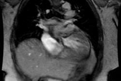
|
Anomalous LCA origin from the right coronary sinus: Coronary CT angiography was performed to assess for coronary artery disease. The patient was found to have an anomalous LCA arising from the right sinus of valsalva. The vessel can be seen to course between the pulmonary trunk and aorta (white arrows). Click the image to view the video clip. |
|
|

REFERENCES:
(1) Ann Thorac Surg 1998; Radke PW, et al. Anomalous origin of the right coronary artery: Preoperative and postoperative hemodynamics. 66: 1444-1449
(2) AJR 2004; Hague C, et al. MDCT of a malignant anomlaous right coronary artery. 182: 617-618
(3) J Nucl Med 2004; De Luca L, et al. Stress-rest myocardial perfusion SPECT for functional assessment of coronary arteries with anomalous origin or course. 45: 532-536
(4) Radiology 2005; Datta J, et al. Anomalous coronary arteries in adults: depiction at multi-detector row CT angiography. 235: 812-818
(5) AJR 2005; Khanna A, et al. Anomalous origin of the left coronary artery from the pulmonary artery in adulthood on CT and MRI. 185: 326-329
(6) Radiographics 2006; Kim SY, et al. Coronary artery anomalies: classification and ECG-gated multi-detector row CT findings with angiographic correlation. 26: 317-334
(7) J Nucl Cardiol 2006; Chhatriwalla AK, et al. An 8-month-old girl with an anomalous left coronary artery from the pulmonary artery complicated by myocardial ischemia after surgical reimplantation. 13: 432-436
(8) Radiographics 2009; Pena E, et al. ALCAPA syndrome: not just a pediatric disease. 29: 553-565
(9) J Nucl Cardiol
2009; van der Jagt
LH, et al. An anomalous RCA with ischemia on
myocardial perfusion imaging. 16: 474-477
(10) AJR 2011; Young PM, et al. Cardiac imaging: part 2, normal,
variant, and anomalous configurations of the coronary vasculature.
197: 816-826
(11) Radiology 2012; Lee HJ, et al. Anomalous origin of the right
coronary artery from the left coronary sinus with an intraarterial
course: subtypes and clinical importance. 262: 101-108
(12) J Cardiovasc Comput Tomogr 2012; Pursnani A, et al. Coronary
CTA assessment of conrary anomalies. 6: 48-59
(13) Radiographics 2012; Shriki JE, et al. Identifying,
characterizing, and classifying congenital anomalies of the
coronary arteries. 32: 453-468
(14) J Cardiovasc Comput Tomogr 2013; Aldana-Sepulveda N, et al. Single coronary artery: spectrum of imaging findings with multidetector CT. 7: 391-399
(15) Radiographics 2017; Agarwal PP, et al. Anomalous
coronary arteries that need intervention: review of pre- and
postoperative imaging appearances. 37: 740-757