Hamman-Rich Syndrome (Acute Interstitial Pneumonia):
View cases of AIP
Clinical:
Acute interstitial pneumonia (AIP) is the preferred term for this autoimmune disorder characterized by rapidly progressive organizing form of diffuse alveolar damage with permeability edema [10]. The condition consists of partially overlapping stages (exudative, proliferative, and ultimately with interstitial pulmonary fibrosis) [10]. AIP can be idiopathic or secondary to some other insult [10].Idiopathic AIP typically affects patients at a younger age (the mean age of affected patients is 50 years [7]), but the condition can be seen at any age [11]. The clinical course is rapidly progressive (over weeks to a few months, as opposed to years). Men and women are affected equally and cigarette smoking does not seem to increase the risk for AIP [7]. Typically a history of a viral like illness exists [7]. Patients present with rapidly progressive dyspnea (over days), hypoxemia, and respiratory failure. Treatment is largely supportive [7] and mortality ranges from 50-100%.
Histologically the hallmark of the disorder is diffuse alveolar damage (DAD)- the term AIP is reserved for patients with DAD of unknown etiology [6]. Secondary AIP is the morphologic basis of ARDS which can be caused by exposure to infectious pathogens, chemical agents, or sepsis/trauma [10].
Histologically, there are three partly overlapping stages- exudative, proliferative, and fibrotic [11]. Diffuse alveolar damage manifests initially as injury to the alveolar lining with widespread sloughing of type I endothelial cells, pulmonary edema, hemorrhage, and hyaline membrane formation [9]. There is also widespread alveolar collapse that leads to severe hypoxemia. A chronic fibrotic phase follows the acute phase if patients survive [6]. Survivors can have significant residual parenchymal abnormalities. AIP is a diagnosis of exclusion with negative bacterial, viral, and fungal cultures, and no evidence of toxic or drug exposure. Some feel that it represents an idiopathic form of ARDS.
X-ray:
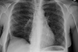
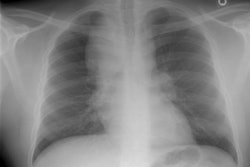
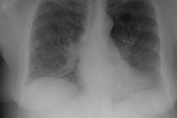
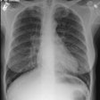
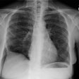
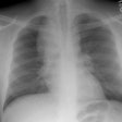
The CXR may initially be normal, but there is usually bilateral, patchy airspace opacifications which spare the costophrenic angles [11]. The cardiac silhouette and vascular pedicle are normal and pleural effusion is uncommon because the condition does not result from cardiac insufficiency [10]. As the disease progresses the lungs tend to become diffusely consolidated. Differential considerations include widespread infection (such as PCP), hydrostatic edema, acute eosinophilic pneumonia, diffuse pulmonary hemorrhage, acute hypersensitivity pneumonitis, and alveolar proteinosis [12].On CT the disorder appears similar to ARDS with bilateral, patchy
ground glass opacities, with or without consolidations, often with
some sparing of individual lobules that produces a geographic
appearance [8]. The abnormalities rapidly progress to diffuse
consolidation with a gravitational gradient particularly affecting
the dependent lung zones with the patient in the supine position
[6,11]. With the patient in the prone position, this gradient will
reverse [11]. Interlobular septal thickening may also be seen (due
to edematous thickening or adjacent alveolar microatelectasis).
In the late phase, traction bronchiectasis and honeycombing are seen and are more severe in the non-dependent portions of the lungs (reflecting a protective effect of atelectasis and consolidation on mechanical ventilation associated damage) [7]. Pleural effusion can be found in up to 30% of patients at CT [3].
REFERENCES:
(1) RadioGraphics 1996;16:1009-1033
(2) AJR 1997; Feb 168 (2): 333-338
(3) Radiology 1999; Johkoh T, et al. Acute interstitial pneumonia: Thin-section CT findings in 36 patients. 211: 859-863
(4) Society of Thoracic Radiology Course Syllabus 2000; Lynch DA, et al. International concensus classification of idiopathic intestitial pneumonias. 52-56
(5) Radiographics 2003; Wittram C, et al. CT-histologic correlation of the ATS/ERS 2002 classification of idiopathic interstitial pneumonias. 23: 1057-1071
(6) Radiology 2005; Lynch DA, et al. Idiopathic interstitial pneumonias: CT features. 236: 10-21
(7) Radiographics 2007; Mueller-Mang C, et al. What every radiologist should know about interstitial pneumonias. 27: 595-615
(8) Radiology 2008; Hansell DM, et al. Fleishner society: glossary of terms for thoracic imaging. 246: 697-722
(9) Radiology 2010; Galvin JR, et al. Collaborative radiologic
and histopathologic assessment of fibrotic lung disease. 255:
692-706
(10) AJR 2013; Nemec SF, et al. Lower lobe-predominant diseases
of the lung. 200: 712-728
(11) AJR 2013; Nemec SF, et al. Noninfectious inflammatory lung
disease: imaging considerations and clues to differential
diagnosis. 201: 278-294
(12) Radiographics 2015; Sverzellati N, et al. American thoracic society-European respiratory society classification of the idiopathic interstitial pneumonias: advances in knowledge since 2002. 35: 1849-1872

