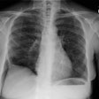
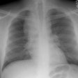
Relapsing Polychondritis:
View cases of relapsing polychondritis
Clinical:
Relapsing polychondiritis is a rare, systemic autoimmune inflammatory disease of unknown etiology which involves the cartilage of the nose, ears (85%), upper respiratory tract (50-70%), and joints. It is probably due to an autoimmune response with antibodies directed against type II collagen [3]. About 30% of cases occur in association with other inflammatory rheumatic, collagen vascular, or vasculitic diseases [2]. The peak incidence occurs between the ages of 40 to 60 years without a sex predilection [2]. Fewer than 10% of cases are seen in children [4]. Caucasions are primarily affected [2]. Laryngotracheal involvement is seen more commonly in women and occurs at presentation in about 10% of patients, but will eventually occur in 50% of patients [7,8]. Involvement of the respiratory tract can affect all cartilage containing portions of the trachea and bronchi and is associated with a poor prognosis (mortality approaches 50%). Nearly 25% of patients will have an associated collagen vascular disease. The disorder is characterized by recurrent episodes of cartilagenous inflammation which eventually leads to fibrous replacement of the damaged cartilage and fixed narrowing of the trachea. Rarely, flaccidity may result. Patients usually present with recurring biauricular and nasal chondritis, as well as inflammatory systemic polyarthritis [4]. Treatment involves steroids to suppress inflammation during periods of disease activity, and dilatation and stenting if strictures have occurred.
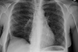
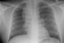
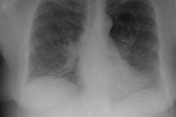
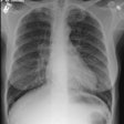
X-ray:
On CT, there is increased airway attenuation. Wall thickening (greater than 2 mm) typically involves the trachea and main bronchi and is usually smooth, diffuse and characteristically spares the posterior membranous wall of the trachea [7,9]. The wall thickening may appear circumferential if the inflammation spreads into the posterior membrane form the adjacent cartilages [9]. Focal tracheobronchial stenoses may also occur [9]. Expiratory collapse (of greater than 50%) of the affected airway is related to loss of cartilagenous support of the airways walls and is best identified dynamic expiratory CT [5,6,8].
Differential considerations for diffuse tracheal narrowing include: Amyloidosis, Sarcoid, tuberculosis, and Wegeners granulomatosis
REFERENCES:
(1) J Thorac Imaging 1995;10(4):236-254 (p.236)
(2) J Thorac Imaging 1992; Tanoue LT. Pulmonary involvement in collagen vascular disease: A review of the pulmonary manifestations of the Marfan syndrome, ankylosing spondylitis, Sjogren's syndrome, and relapsing polychondritis. 7 (2): 62-77 (Review)
(3) Radiographics 1998; Meyer CA, White CS. Cartilagenous disorders of the chest. 18: 1109-1123
(4) AJR 2001; Luckey P, et al. Diagnostic role of inspiration and expiration CT in a child with relapsing polychondritis. 176: 61-62 (No abstract available)
(5) AJR 2001; Marom EM, et al. Diffuse abnormalities of the trachea and main bronchi. 176: 713-717 (No abstract available)
(6) AJR 2002; Behar JV, et al. Relapsing polychondritis affecting the lower respiratory tract. 178: 173-177
(7) Radiographics 2002; Nonneoplastic lesions of the tracheobronchial wall: radiologic findings with bronchoscopic correlation. 22: S215-S230
(8) Radiology 2006; Lee KS, et al. Relapsing polychondritis:
prevalence of expiratory CT airway abnormalities. 240: 565-573
(9) Radiology 2012; Yamamoto AK, Babar JL. Case 184: ulcerative
tracheobronchitis. 264: 609-613

