CHICAGO - Cardiac MRI has uncovered the unusual ability of free divers -- who descend hundreds of feet into the ocean while holding their breath -- to develop "compensatory mechanisms" to recover heart function within minutes of breathing again.
The researchers from Germany, who presented the study at RSNA 2015 on Tuesday, likened the changes in the divers' systolic heart function during the breath-holds to patients with systolic heart failure. The temporary suspension of breathing is known as apnea.
 Dr. Jonas Dörner.
Free diving can be a very dangerous sport, as divers hold their breath for prolonged periods of time in deep waters, experiencing massive water pressure and physiological changes.
"This study illustrates the known phenomena that prolonged breath-holding resulted in progressive left-ventricular enlargement with an increase of left-ventricular stroke volume, which at the beginning compensated the progressive deterioration of left-ventricular systolic function," said lead author Dr. Jonas Dörner, a radiology resident at the University Hospital of Cologne. "It is a similar pattern, as it occurs in the failing heart or during endurance exercise in athletes."
Free-diving enthusiasts
One reason for the study is the avid participation in free diving by co-author Dr. Lars Eichhorn, from the department of anesthesiology and intensive care medicine at the University Hospital of Bonn. Eichhorn is a free-diving instructor and a member of the managing board of the German Society for Diving and Hyperbaric Medicine.
The study also is a follow-up to previous research by Dörner, Eichhorn, and colleagues.
"In our first study, we could visualize prolonged oxygen supply in the brain. This is most likely caused by elevated cerebral blood flow," Dörner told AuntMinnie.com. "Nevertheless, we could only speculate about the effects on our cardiovascular system. Therefore, in this study, we focused on mechanisms that could explain our results."
The group examined the simulated effects of free diving on the cardiovascular systems of 17 elite divers (15 men and two women) from Germany and Austria, who ranged in age from 23 to 58 years.
The subjects underwent a 1.5-tesla cardiac MRI to assess left-ventricular end-diastolic volume, end-systolic volume, ejection fraction, fractional shortening, and other measures. A second MRI scan was performed to measure blood flow in both common carotid arteries using phase contrast imaging.
The average apnea, or breath-hold time, was 299 seconds for the cardiac MRI portion of the study and 279 seconds for the carotid artery MRI exam. The maximum breath-hold during the MRI scans was eight minutes and three seconds, or 483 seconds.
Compared with other modalities, cardiac MRI has several advantages for imaging heart function and functional alterations over time, Dörner said.
"First, cardiac MRI is considered the gold standard for the assessment of cardiac function," he said. "Second, it provides the most robust technique with a low inter- and intraobserver variability and is therefore well-suited for comparison studies. Ultimately, being a nonionizing technique, cardiac MRI can be used repeatedly."
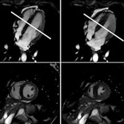
 Cardiac MRI shows normal left-ventricular function in the standard horizontal long axis (upper two images) and corresponding short axis (lower two images) in diastole (left) and systole (right) at the midventricular section of the heart (white line). Images courtesy of RSNA and Dr. Jonas Dörner.
The MRI exams allowed the researchers to observe the cardiovascular changes involved in the diving reflex in real-time. During apnea, the amount of blood flowing to the brain through the carotid arteries increased and then leveled off.
At the beginning of the breath-hold period, the heart pumped more strongly than when the heart was at rest, the researchers found. Over time, the heart dilated and began to struggle, and by the end of the apnea period, the divers' heart function began to fail.
At that end point, not enough blood is being pumped to the brain, Dörner explained, and the heart is unable to pump against the high resistance of the blood vessels.
And while the changes in the divers' systolic heart function during apnea are similar to patients with systolic heart failure, the major finding is that the divers' heart function recovered within minutes of breathing again. This suggests that the elite free divers develop mechanisms that help them adapt to the cardiovascular changes that occur during apnea.
As to whether there is any evidence of potentially permanent physical damage to trained free divers over a long period of participation in the sport, the research so far has only shown the "tip of the iceberg," Dörner said. "In this moment, we just focused on immediate changes of the cardiac function. Long-term effects have to be answered in further studies."
He cited one study on the long-term effects on blood pressure in elite divers; the group did not show a significantly greater incidence of hypertension.
"Up to now, there is no specific evidence that there may be potentially permanent physical organ damage," Dörner added.