MONTREAL - Results from two MR imaging techniques have Canadian researchers challenging the thinking that treatment for pediatric brain tumors damages myelin, according to a study presented Wednesday at the International Society for Magnetic Resonance in Medicine (ISMRM) annual meeting.
Using diffusion-tensor imaging (DTI) and magnetization transfer imaging (MTI), the researchers found decreased fractional anisotropy and increased diffusivity, but no significant differences in magnetic transfer. The findings would suggest that the pediatric patients' treatment might adversely affect fiber organization rather than myelin structure.
"The current study challenges the paradigm that myelin injury is a consequence of pediatric brain tumors and their treatment," wrote lead author Logan Richard, MRI technician, and colleagues at the Hospital for Sick Children in Toronto. "Magnetization transfer ratio should be used as an adjunct to DTI in order to refine interpretation of data within future research in this patient population."
Pediatric cancers
Tumors of the central nervous system are the second most common form of pediatric cancer, accounting for approximately 25% of all cases. Treatment options include surgery and/or radiation, with strategies dependent upon disease dissemination, tumor size, and diagnosis.
While treatment protocols have advanced over the years, the young patients are exposed to therapeutic regimens at a sensitive time during their physical development. The effects can leave survivors with compromised white matter cognitive deficiencies.
Previous research into white matter microstructure in pediatric brain tumor survivors has focused on diffusion-tensor MR imaging (DTI-MRI). However, results from DTI can be altered by several factors, such as edema and inflammation.
"The nonspecificity of DTI data can, therefore, make interpretation of white matter microstructure challenging," said graduate student Elizabeth Cox, who presented the results at ISMRM. "In order to mitigate the imprecision of DTI, MTI can also be employed to provide a more specific measure of white matter myelin in pediatric brain tumor survivors."
What is MTI?
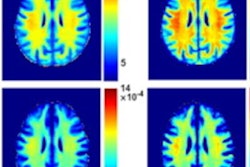
MTI measures signals from macromolecular surroundings. In a white matter environment, the predominant macromolecular structure is that of lipids and myelin. Signal acquired before a magnetization transfer (MT) pulse is computed with the signal acquired after an MT pulse, she explained.
"The MT signal acquired during 'MT on' is impacted by myelin, which creates a lower signal than 'MT off,' if the myelin is healthy," Cox added. "This signal is used to compute a magnetization transfer ratio (MTR). A higher MTR indicates greater myelin integrity."
The researchers recruited 50 pediatric brain tumor survivors (mean age, 12.4 years) and 45 healthy control subjects, who matched for age and gender, from the Hospital of Sick Children. Among the surviving patients were 18 children who were treated with surgery only and 32 children who underwent radiation with or without surgery. Treatment ranged from reduced (2340 cGy) to standard (3060 to 3940 cGy) doses of radiation to the head and spine, as well as a radiation boost to either the tumor bed or entire posterior fossa.
For the study, the subjects were divided into three groups: pediatric patients who received surgery only, patients who were treated with surgery and radiation, and the healthy controls.
MRI exams were performed on a 3-tesla scanner (Siemens Healthineers), with protocols that included a T1-weighted axial 3D magnetization-prepared rapid gradient-echo (MPRAGE) and a diffusion-weighted single shot spin-echo DTI sequence, as well as calculation of an MTR.
Researchers' hypotheses
From the protocols, Cox and colleagues used DTI and MTI to observe white matter integrity in tumor survivors and compared the efficacy of the two techniques.
They hypothesized the following:
- Using DTI, there would be areas of reduced fractional anisotropy and increased radial diffusivity in pediatric tumor patients, compared with healthy controls.
- Using MTI, there would be lower MTR in patients relative to controls.
- When comparing DTI and MTI, there would be lower MTR in fewer regions than found with DTI in tumor patients.
"Our primary analyses investigated whether there was a significant difference between healthy controls and all patients across all DTI indices and MTR," Cox said.
Outcome comparisons
The first analysis confirmed the first hypothesis with a statistically significant decrease in fractional anisotropy in both patient groups, compared with the healthy subjects (t = 2.18; p = 0.0012). The researchers also noted statistically significant increases in axial diffusivity (t ≥ 2.6; p < 0.03), radial diffusivity (t = 2.14; p < 0.001), and mean diffusivity (t = 1.97; p < 0.001) among both patient groups, compared with the healthy controls.
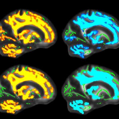
 MR images compare fractional anisotropy (FA) and radial diffusivity (RD) between the surgery (yellow) and radiation (blue) patient groups and healthy controls. Colored areas indicate significant brain regions showing decreased FA and increased RD within patient groups. Mean FA skeleton is shown in green. Images courtesy of ISMRM.
Brain regions among the surgical patients showed widespread reduction of fractional anisotropy -- compared with healthy controls -- across the anterior, posterior, and bilateral regions.
In addition, the researchers noted no statistically significant differences in MTR in comparisons between the surgical group (p = 0.32) and the radiation therapy group (p = 0.33) and healthy controls.
"As a reminder, decreased fractional anisotropy and increased diffusivity are thought to be reflective of less organized white matter microstructure, and decreased MTR is thought to reflect compromised white matter myelin integrity," Cox added. And, given that the team found no difference in MTR, it would suggest that myelin integrity was not affected.
Based on the numbers, the researchers wrote in their summary that "myelin itself may not be affected by pediatric brain tumors and their treatment. Rather, it appears that treatment could impact other factors that may disrupt fiber organization, including axonal injury, axonal density, fiber packing, and fiber orientation."