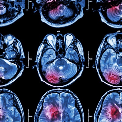
A 7-minute MRI protocol can now play a central role in the clinical management of acute ischemic stroke (AIS) patients, award-winning researchers have reported at ECR 2023.
"The sophisticated MRI protocol can contribute both to the faster reperfusion and to accurate diagnosis," noted Daisuke Oura and colleagues from the Department of Radiology, Otaru General Hospital, Otaru, Japan. "Recently, the importance of evaluation of brain tissue using imaging examination has grown."
The group received one of seven magna cum laude awards for its electronic poster award. The three European winners came from Spain, it was revealed on the opening day of the congress in Vienna. There was another magna cum laude winner from Japan, and the other two came from Latin America.
How to shorten exam time
Oura, who is a doctoral student at Hokkaido University in Sapporo, and colleagues explained that AIS due to large vessel occlusion often leads to serious disability, and the time taken for reperfusion is the most important factor to obtain a better clinical outcome. Premechanical thrombectomy imaging using CT or MRI must be shortened, they added.
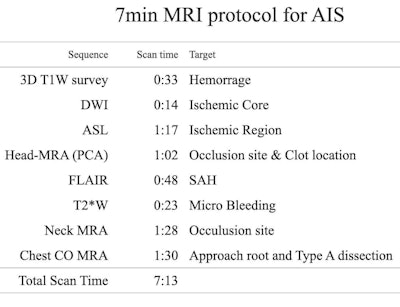
The authors have refined their MRI protocol and developed a rapid 1-min phase contrast angiography (PCA), after handling more than 600 cases of suspected AIS cases using a 3-tesla Ingenia machine (Philips) with a 32-channel head coil. This protocol included the following sequences; diffusion-weighted imaging (DWI), arterial spin labeling (ASL), head MR angiography (MRA), FLAIR, T2*WI, neck MRA, and chest coronal MRA.
 Details about the 7-minute MRI protocol. SAH = subarachnoid hemorrhage. Tables courtesy of Daisuke Oura et al and presented at ECR 2023.
"AIS patients often show motion artifacts due to ischemia of a large cerebral region. Conventional MRA using time-of-flight (TOF) is sensitive to motion artifacts, which degrades image quality," they noted. "Additionally, a scan of under 60 seconds loses image quality in time-of-flight. We overcame these disadvantages by optimizing PCA for cerebral arteries."
The researchers performed a visual evaluation for image quality compared with TOF-MRA. Images from 10 healthy volunteers were studied by two experienced technologists using the five-point scale method. The rapid 1-minute PCA delivered a significantly higher image quality and a shorter scan time. The shortest repetition time provided the robustness for motion artifact.
The black-blood image was created by the image subtraction between the source image of PCA, using the DEPICT (Depicting of blood clot and MRA using Phase contrast angiography with an Image Calculation for Thrombectomy) technique.
"Generally, rapid chest MRA using 3-tesla MRI tends to become poor quality due to B1 nonuniformity or incomplete fat suppression. We developed the rapid MRA without any prepulse and fat suppression," the authors wrote.
Repetition time and echo time were set as short as possible, without any prepulse in a 2D single-shot turbo field echo sequence. The soft tissue is suppressed by the saturation effect, and the in-flow effect is maximized; this sequence is called simple T1-TFE, they stated.
"Actual manipulation time is one of the important factors to achieve faster reperfusion," they continued. "We compared the manipulation time by 10 radiographers between our MRI and CT protocol in the phantom study. 10 technologists imaged the phantom with both MRI protocol and CT protocol for AIS."
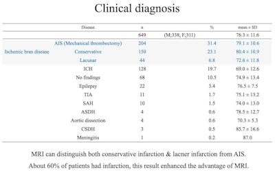
 Clinical diagnosis of over 600 cases of suspected AIS. Over 60% of patients had ischemic disease. MRI had higher sensitivity and specificity than CT.
Approximately 60% of patients had ischemic brain diseases such as AIS, conservative infarction, and lacunar infarction. The findings support the use of MRI as first choice for suspected cases of AIS.
"Patients with diseases mimicking AIS such as epileptic seizure and transient ischemic attack were also quickly screened," the authors concluded. "The results show the clinical usefulness of our 7-min MRI protocol."
The same Otaru group published an article in the November 2022 edition of Magnetic Resonance Imaging about the simultaneous depiction of clot and MRA using 1-min phase contrast angiography in acute ischemic patients.
Top prize-winners in Vienna
The following are the six other ECR 2023 magna cum laude award winners:
- Videofluoroscopic evaluation of velopharyngeal insufficiency: a quick guide for residents. S. J. Valdes Bañuelos et al, Leon, Mexico.
- Beyond acute cholecystitis: Unravelling step-by-step imaging findings of its main complications. D. Herrán De La Gala et al, Santander, Spain.
- Malignant transformation of benign gynecologic diseases: Wide Spectrum of Clinical and Imaging Manifestations, Differential Diagnosis and Pitfalls. M. Takeuchi et al, Tokushima, Japan.
- Extrapulmonary tuberculosis: imaging features beyond the chest. I. Díaz Mediavilla et al, Bilbao, Spain.
- Total ankle replacement. Radiological review of imaging clues in pre- and post-operatory assessment of ankle arthroplasty. F. J. Azpeitia Arman et al, Madrid, Spain
- Radiological Patterns of the Thoracic Synovial Sarcoma: One of the rarest locations! R. Pareja Maldonado et al, Lima, Peru


