VIENNA - Radiation doses to the breast can be cut by nearly 40% using a new organ-based dose modulation technique in combination with a noise-optimized image reconstruction method.
In a presentation at the 2010 European Congress of Radiology (ECR), researchers from Siemens Healthcare in Erlangen, Germany, said the two techniques must be used together if the resulting images are to remain readable.
Modern scanners offer several ways to modify the tube current; for example, by modulating the mAs during the systolic phase of coronary CT angiography, or by varying tube current based on attenuation, noted Bernhard Schmidt, Ph.D.
Unfortunately, existing dose-reduction methods may not be good enough. Publication of new tissue weighting factors in the most recent International Commission on Radiological Protection (ICRP) guidelines (Publication 103, 2007) revealed that organ doses, particularly to the breast, may be more important than previously thought.
"In the weighting factors for breast tissue, there was a significant change from the old rating factors from the old weighting of 0.05 to the new weighting of 0.12, an increase of more than a factor of two," Schmidt said. "The risk for breast tissue is significantly higher than previously assumed, so we have to look into techniques to reduce the dose to the breast tissue."
The study assessed organ-based tube-current modulation using the method to examine both dose and image quality, including the assessment of a dedicated noise-optimized reconstruction technique (DRT).
The dose-reduction scheme, dubbed xCare, reduces breast dose by reducing tube current during each rotation for anterior projections down to 10% of normal, while maintaining the same total mAs per rotation as a nonmodulated scan and, therefore, the same volume CT dose index (CTDIvol) calculation, Schmidt said.
By itself, however, the dose-reduction technique produces a significant increase in image noise due to the greatly reduced signal from the low-dose anterior projection.
"The reason is that the weight of each ray is independent of the angle-specific tube current," he said, "and this, in the end, leads to a noise increase in our data, and therefore we propose a dedicated noise-optimized reconstruction technique."
Because the modulation technique isn't based on attenuation, projections with high tube current must receive during reconstruction greater weighting toward the final image reconstruction than the projections acquired with low tube current -- which is precisely the job of the DRT technique, which is applied only to the anterior projections, Schmidt noted.
The researchers measured doses using an anthropomorphic phantom with breast attachments into which 14 thermoluminescence detectors were placed.
For circular phantoms, they found that noise increased in the periphery of the breast without DRT by 1%, 17%, and 57%, when reducing tube current in the low dose plateau down to 50%, 30%, and 10%, respectively.
However, image quality is restored once the DRT is applied, by a factor of about two at 10% of normal dose for the anterior projection. Similar results were obtained in tests of the anthropomorphic phantom, where "noise is reduced to the same levels as before the tube-current reduction," Schmidt said.
Noise levels without and with DRT
|
||||||||||||||||||||||||||||||||||||||||||
Phantom results show that application of the DRT restored low noise levels obtained with full-dose scans.
As for dose, based on readings of the thermoluminescence detectors placed in the anthropomorphic phantom, reductions of about 30% to 40% were achieved.
Organ dose: Breast
|
Thermoluminescence detector measurements show dose reductions of up to 36% using the xCare organ-based dose-reduction method on reduced tube-current anterior projections.
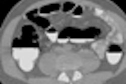
Finally, slides of several patients scanned with the method showed no reduction in image quality before and after the dose reductions, according to Schmidt.
"Organ-based tube-current modulation allows us to reduce dose to the breast by 30% to 40%," he concluded. "We need a dedicated reconstruction technique; otherwise, we would see a significant increase in noise in our data. The goal, of course, has to be a combination of organ-based tube-current modulation with the z-axis tube-current modulation to minimize the dose to the breast tissue."
The technique is also being applied to CT-guided fluoroscopy, where it reduces dose both to patients and to the hand of the fluoroscopy operator, Schmidt said in response to a question from the moderator.
By Eric Barnes
AuntMinnie.com staff writer
March 8, 2010
Related Reading
Inexperienced readers benefit most from breast US CAD, March 5, 2010
Radiation exposure increases risk of breast cancer, March 4, 2010
CARS report: Novel conebeam CT cuts breast dose, improves accuracy, June 29, 2009
ECR delivers new findings on digital breast tomosynthesis, March 7, 2009
Italian study backs tomosynthesis over DR for lung pathology, February 18, 2009
Copyright © 2010 AuntMinnie.com