CHICAGO - Dark-field x-ray may be more effective than conventional radiography for diagnosing and staging pulmonary emphysema, according to research presented November 27 at RSNA in Chicago.
A group at the Technical University of Munich (TUM) in Germany tested the technique in a head-to-head study versus conventional x-ray for diagnosing and staging pulmonary emphysema. They found dark-field chest x-ray outperformed conventional radiography.
"Compared to conventional chest radiography, dark-field chest radiography provides improved capability for identification and staging of emphysema," said presenter Theresa Urban, a 2022 RSNA 2022 trainee research prize winner.
Dark-field chest x‑ray is an experimental technique based on modifying existing x‑ray scanners. The approach uses interferometers to visualize the scattering of x‑rays in the alveoli of the lungs, with higher numbers of interfaces leading to stronger dark-field signals.
In this study, the group enrolled 88 patients with either no lung impairment or any stage of emphysema. CT scans served as a visual reference, with emphysema absent in 36 patients, trace disease in 21, mild in 11, moderate in nine, confluent in seven, and advanced in four patients.
Visual assessment of images was performed by clinical radiologists, with three readers categorizing conventional x-ray images and dark-field chest x-ray images as no emphysema, mild, moderate or severe.
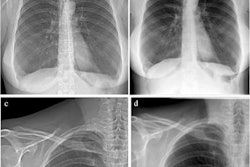
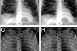
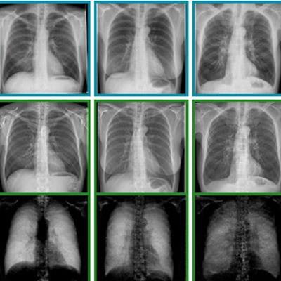
 Exemplary radiographs and dark-field chest x-ray images from the study. Image courtesy of Theresa Urban.
Dark-field images showed a distinct decrease of signal strength with emphysema severity, as alveoli are denser in these patients, according to the findings. Readers were significantly better able to identify emphysema with images from the dark-field prototype, achieving an area under the curve (AUC) of 0.85 (p < 0.05) compared with conventional images (AUC, 0.74).
In addition, while ratings of adjacent emphysema severity groups with conventional radiographs were only different for trace and mild emphysema, ratings based on images from the dark-field prototype were different for trace and mild, mild and moderate, and moderate and confluent emphysema.
"Readers were better able to detect emphysema with images from the dark-field chest radiography system," Urban said.
This was the first head-to-head study pitting the new technique against conventional chest x-rays, and more work is planned, Urban told AuntMinnie.com. The approach yields both attenuation and dark-field x-rays in a single acquisition, and these images may be combined, which could offer even more diagnostic accuracy, she added.
"Dark-field chest radiography demonstrates high diagnostic value for lung assessment," Urban concluded.