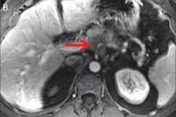
Contrast-enhanced ultrasound is more accurate than conventional ultrasound for distinguishing between focal pancreatitis and pancreatic cancer, according to a study published in the March issue of the Journal of Ultrasound in Medicine.
It can be hard to tell the two diseases apart, and the findings suggest that contrast-enhanced ultrasound shows promise as a more effective tool in the clinical arsenal, wrote the group led by Yanjie Wang, PhD, of Peking University Cancer Hospital and Institute in Beijing.
"As contrast-enhanced ultrasound can reflect the microvessels with real-time and dynamic methods, [its] diagnostic performance ... is similar to that of contrast-enhanced CT for diagnosing pancreatic carcinoma and focal pancreatitis," the researchers wrote.
Detecting the difference
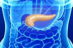
Focal pancreatitis can manifest clinical symptoms similar to those of pancreatic cancer, such as jaundice, weight loss, and abdominal masses. It is often treated with a Whipple procedure, otherwise known as a pancreaticoduodenectomy -- a surgical procedure that removes the head of the pancreas, the first part of the small intestine, the gallbladder, and the bile duct. This can have far-reaching negative effects on patients, especially if the diagnosis is incorrect, Wang and colleagues wrote (J Ultrasound Med, March 2018, Vol. 37:3, pp. 551-559).
"As the treatment for these diseases are completely different, a misdiagnosis might result in the unnecessary traumatic resection of benign lesions or missing the opportunity for radical resection of malignant lesions," they wrote.
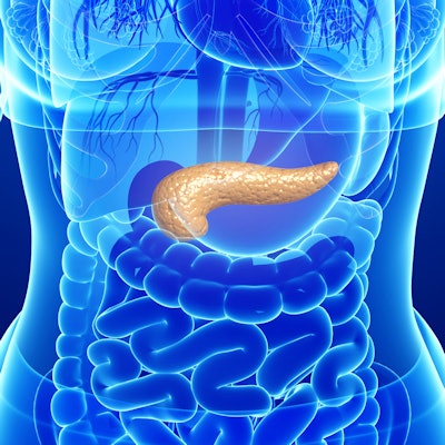
Conventional ultrasound has long been used as a first tool for diagnosing pancreatic lesions. But contrast-enhanced ultrasound offers a better way to characterize them because it can visualize microvascular perfusion in real-time, according to the researchers.
For the study, they included 136 solid pancreatic lesions diagnosed between January 2010 and February 2016. Of these cases, 25 were focal pancreatitis (mean diameter, 4.2 cm), 86 were pancreatic carcinoma (mean diameter, 3.8 cm), and 25 were other diseases (such as neuroendocrine tumors, solid pseudopapillary tumors, lymphomas, and myofibroblastomas; mean diameter, 3.3 cm).
All patients underwent conventional and contrast-enhanced ultrasound exams. Contrast-enhanced exams were divided into early phase (10-30 seconds after injection) and late phase (30-120 seconds after injection). Two experienced ultrasound physicians analyzed the contrast-enhanced exams, categorizing them into five patterns:
- Isoenhancement
- Isoenhancement with focal hypoenhancement
- Centripetal enhancement
- Hypoenhancement
- Hyperenhancement
The first two patterns, in both early and late contrast phases, were considered diagnostic for focal pancreatitis, the group wrote.
The researchers then compared the diagnostic efficiency of contrast-enhanced ultrasound exam patterns versus conventional ultrasound exam patterns for differentiating focal pancreatitis from pancreatic cancer, calculating sensitivity, specificity, accuracy, positive predictive value (PPV), and negative predictive value (NPV).
Wang and colleagues found statistically significant agreement for contrast enhancement patterns between the two study readers (p < 0.05). In addition, with contrast-enhanced ultrasound, focal pancreatitis showed isoenhancement in 32% of the cases and isoenhancement with focal hypoenhancement in 40% of the cases in both the early and late phases.
Compared with isoenhancement in both the early and late phases, the diagnostic sensitivity of isoenhancement or isoenhancement with focal hypoenhancement in both the early and late phases was greatly increased for diagnosing focal pancreatitis, from 32% to 72%, the authors noted. Diagnostic specificity, accuracy, PPV, and NPV showed no significant differences.
Pancreatic cancer showed centripetal enhancement in the early phase of the contrast-enhanced ultrasound exams and hypoenhancement in the late phase in 33.7% of cases, and hypoenhancement in both the early and late phases in 55.8% of cases.
Contrast-enhanced ultrasound proved more accurate in distinguishing between the two diseases than conventional ultrasound, at 85.6% versus 49.5% (p < 0.001), Wang and colleagues found.
| Conventional US vs. contrast US for pancreatitis and pancreatic cancer | |||
| Parameter | Conventional ultrasound | Contrast-enhanced ultrasound | p-value |
| Accuracy | 49.5% | 85.6% | < 0.001 |
| Undetermined result | 45.9% | 5.4% | < 0.001 |
| Error | 4.5% | 9% | 0.322 |
"The rate of an undetermined [result] decreased greatly when contrast-enhanced ultrasound was used for diagnosing focal pancreatitis and pancreatic carcinoma than that of conventional ultrasound ... [and] diagnostic accuracy increased greatly," they noted. "The diagnostic error had no statistically significant differences."
Better tool
Contrast-enhanced ultrasound improves the diagnosis of pancreatic lesions, and because it can reflect the microvessels with real-time and dynamic methods, its diagnostic performance is similar to that of contrast-enhanced CT -- without the radiation, the researchers wrote.
"The application of contrast-enhanced ultrasound greatly increased the diagnostic accuracy over conventional ultrasound for differentiating focal pancreatitis from pancreatic carcinoma," they concluded.