Korean radiologists have developed a new MRI grading system to more accurately assess the degree of cervical canal stenosis in patients with back problems, according to a study published in the July issue of the American Journal of Roentgenology.
With the system, the grade of stenosis positively correlated with the number of patients who had neurologic deterioration indicating myelopathy, the authors wrote. The grade also correlated with the number of patients who had either undergone surgery or were considered candidates for an operation.
"This finding indicates that our grading system conveys clinically significant information and that it may correlate well with the recommended indications for surgical treatment, although further studies are mandatory," concluded lead author Dr. Yusuhn Kang, from the department of radiology at Seoul National University, and colleagues (AJR, July 2011, Vol. 197:1, pp. W134-W140).
 |
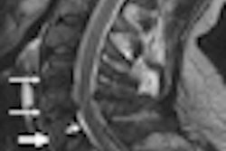
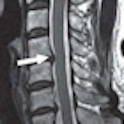
| Image of a 61-year-old woman with cervical canal stenosis. Sagittal T2-weighted fast spin-echo image shows grade 2 stenosis at C4-5 level (arrow). Spinal cord is compressed and deformed but shows no signal changes. Grade 1 stenoses were also seen at C5-6 and C6-7 levels. Image courtesy of AJR. |
Kang and colleagues advocated that a standardized grading system should be a "prerequisite for the comparison of data from different investigations and for the improvement of communication between radiologists and clinicians."
Previous research
The current study is based on research by a German group from Christian-Albrechts-University Kiel (American Journal of Neuroradiology, 1998, Vol. 19, pp. 1763-1771).
Dr. Claus Muhle and colleagues created a grading system to evaluate four stages of degenerative disease of the cervical spine in 81 patients who had undergone MRI. Spinal stenosis was classified for each segment on a four-point grading system in which 0 was normal, 1 meant partial obliteration of the anterior or posterior subarachnoid space, 2 was complete obliteration of the anterior or posterior subarachnoid space, and 3 was cervical cord compression or displacement.
Kang and colleagues used T2-weighted sagittal MR images to classify cervical canal stenosis into four grades. Grade 0 meant there was no canal stenosis, grade 1 signified subarachnoid space obliteration exceeding 50%, grade 2 indicated spinal cord deformity, and grade 3 indicated spinal cord signal change.
The retrospective study reviewed cervical spinal MRI scans of 100 patients older than 60 years. Kang's group chose subjects older than 60 based on previous research suggesting that specimens from cadavers in this age group had significantly narrower canals than specimens from younger donors. "This helped to eliminate a majority of MRI scans with negative findings," they wrote.
Among the participants, 50 had undergone cervical spinal MRI exams at Seoul National University between January and September 2009. Another 50 subjects had received cervical spine MR imaging at other hospitals.
The researchers then excluded 18 patients from the study because of a history of trauma, previous spinal operations, or histologically proven or suspected tumors. The final study population included 37 men and 45 women, with a mean age of 65.2 years, ranging from 60 to 86 years.
Imaging protocol
At Seoul National University, patients were scanned on either a 1.5-tesla MRI system (Gyroscan, Philips Healthcare) with a head-and-neck coil or a 3-tesla scanner (Intera Achieva, Philips) with a 16-channel neurovascular coil. All patients were imaged in the supine position. MRI exams from other facilities were performed with various MRI scanners using different protocols.
Six radiologists, three of whom had musculoskeletal experience, independently interpreted the images for cervical canal stenosis at three sequential levels (C4-5, C5-6, and C6-7) and were blinded to the patient's history and age.
Intraclass correlation coefficient (ICC) was used to judge interobserver and intraobserver reliability of the new MRI grading system. The researchers adapted a scale from previous research in which ICC values of less than 0.4 indicated poor reproducibility, values of 0.4 to 0.75 indicated fair-to-good reproducibility, and values greater than 0.75 represented excellent reproducibility.
Study results
Kang and colleagues found "moderate-to-substantial" agreement, ranging from 63% to 64%, among the six readers in terms of distinction among the four individual grades. There was a slightly higher level of agreement -- 79% to 85% -- for the presence of central canal stenosis.
Agreement was even greater, at 81% to 85%, in classifying cases as either insignificant stenosis (grades 0 and 1) or significant stenosis (grades 2 and 3). The best agreement -- 92% to 95% -- was obtained for the presence of signal change in a patient's spinal cord.
The average agreement among the three more experienced radiologists did not greatly differ from the average agreement of all six readers, the authors found. Given that the average agreement among all six readers showed little difference, they concluded that the system "is easy to learn, even for the inexperienced radiologists."
As for interobserver agreement as calculated by ICC, values ranged from 0.716 to 0.802, which reflected good-to-excellent agreement. Overall intraobserver agreement was considered excellent at 0.768, they wrote.
Kang and colleagues cited several limitations of the study, including how its retrospective nature may have limited the correlation between the stenosis grade and patient symptoms. In addition, because much of the work was conducted at just one facility, "further multicenter studies are required," they noted.