Full-field digital mammography (FFDM) does not improve upon the performance of film-screen mammography (FSM), at least in terms of the percentage of missed interval and screening-detected cancers, Norwegian researchers reported in a study published online June 14 in Radiology.
In the retrospective study evaluating the percentages and mammographic features missed by FFDM and FSM in the Norwegian Breast Cancer Screening Program (NBCSP), the researchers did not identify a significant difference between the two methods in the percentages of missed interval and screening-detected cancers. However, they did find that cancers missed at FFDM tended to have different mammographic features than those missed with the analog method.
"The use of FFDM has not reduced the challenge of missed cancers," wrote study author Dr. Solveig Hoff, of Aalesund Hospital, and colleagues.
Studies have shown that between 1% and 35% of both interval and screening-detected cancers in patients screened at FFDM can be classified as missed in retrospective reviews of prior mammograms. Because the percentage of missed cancers is a quality measure of a screening program -- and should be kept as low as possible -- systematic review of missed cancers, including prior and diagnostic mammograms, is an important learning tool for radiologists, according to the researchers (Radiology, June 14, 2012).
While FFDM is gradually replacing FSM as a screening tool for breast cancer, there is limited knowledge about the percentage and characteristics of missed cancers in women examined with FFDM, according to the study team.
As a result, Hoff's group compared the percentages and mammographic features of cancers missed at FFDM and film-screen mammography in women who participated in NBCSP between 2002 and 2008. NBCSP offers biennial mammographic screening to women ages 50 to 69; radiologists perform independent double reading with consensus as well as dual-view screening for both initial and subsequent examinations.
The study included 49 cases of interval cancer (three [ductal carcinoma in situ, or DCIS] and 46 invasive) from 18,239 FFDM examinations, and 86 screening-detected cancers (13 DCIS and 73 invasive) from 16,888 FFDM examinations. The comparison group included 81 interval cancer cases (five DCIS and 76 invasive) from 36,271 FSM examinations, and 123 screening-detected cancers (12 DCIS and 111 invasive) from 30,765 FSM examinations.
The researchers found that the percentages of interval and screening-detected cancers missed at FFDM and FSM did not differ significantly.
Missed cancers: FSM vs. FFDM
|
Different misses
The team did find, however, that the types of cancers missed differed between the two modalities:
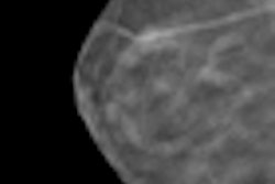
- Asymmetry was present in 27% of the cancers missed at FFDM and in 10% of those missed at FSM.
- Calcifications were observed in 18% of the cancers missed at FFDM and in 34% of those missed at FSM.
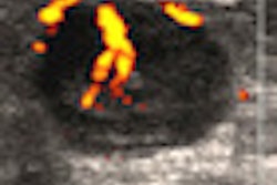
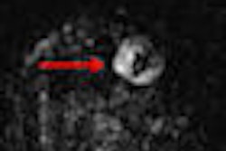
- Average mammographic tumor size of missed cancers manifesting as masses was 10.4 mm at FFDM and 13.6 mm at FSM.
Masses found at FFDM had a significantly smaller mammographic size than masses at FSM. This trend was evident in both prior mammograms of missed cancers and in diagnostic mammograms of screening-detected cancers, according to the authors.
Furthermore, the group discovered trends toward a lower percentage of masses with spiculated margins and a higher percentage of masses with indistinct margins at FFDM compared to FSM.
"These findings can be explained by higher contrast resolution at FFDM than at [film-screen mammography], which makes it easier to distinguish a mass from the surrounding glandular tissue," the authors wrote. "The ability to optimize FFDM images by using postprocessing procedures might further influence the perception and categorization of a small mass."
The authors noted that they expected FFDM to yield a higher percentage of malignancies with calcifications at diagnostic examinations of screening-detected cancer and a corresponding lower percentage in prior mammograms of missed cancer.
"Malignant calcifications are often associated with DCIS and this also corresponds with our finding of a tendency toward a larger proportion of DCIS in screening-detected cancer at FFDM than at [film-screen mammography]," they wrote.
Individual reader performance may have contributed to the higher percentage of missed cancers with asymmetry at FFDM, according to the researchers.
"The high contrast resolution in digital images, the possibility of postprocessing the images in soft-copy reading, and the large size of the computer monitors may have increased reader attention to small details on magnified views at the expense of the overview needed to compare differences in the mammographic patterns between the two breasts," the authors wrote. "To prevent pitfalls in FFDM reading, procedures should be implemented that emphasize the importance of viewing both standard and magnified views in a structured way."
Further studies exploring the mammographic features of missed cancers in FFDM are needed, the authors noted. Furthermore, "individual site reviews of interval and screening-detected cancers are important tools in the ongoing quality assurance of mammographic interpretations," they wrote.