Using diffusion-tensor MRI (DTI-MRI), researchers have discovered significant differences in early brain development in high-risk infants who later develop autism, compared with those who do not develop the disorder, according to a study published online February 17 in the American Journal of Psychiatry.
The results suggest that autism develops over time during infancy, raising the possibility that targeted intervention could interrupt disease progression, said lead study author Jason Wolff, PhD, a postdoctoral fellow at the Carolina Institute for Developmental Disabilities (CIDD) of the University of North Carolina.
The preliminary findings may be a significant first step toward developing a biomarker that could be used to help diagnose autism, he noted.
Autism development
Previous studies have found that siblings of children with autism are at higher-than-average risk for developing the disorder, Wolff and colleagues wrote. Behavioral features of autism begin to emerge at approximately 12 months of age after a period of relatively normal postnatal development. Early studies also have shown that MRI can be used to detect significantly larger than normal brain volume in 2- and 3-year-old children with autism.
"Taken together, findings from the behavioral studies and the [MRI] studies of brain and head size growth in high-risk infant siblings suggest that the latter half of the first year of life is a pivotal time for both brain changes and symptom onset in infants later diagnosed with [autism]," the authors wrote.
Therefore, Wolff and colleagues began a prospective study using DTI-MRI to gauge the development of white-matter fiber tracts in 92 children at age 6, 12, and 24 months. The children were considered at risk for autism because they had an older brother or sister with the affliction. The researchers focused on white-matter fiber tracts because they act as pathways to connect different brain regions.
The study included all high-risk infants who received DTI-MRI scans at six months and behavioral assessments at 24 months, as of June 2011. Symptoms of autism spectrum disorders (ASDs) were measured at 24 months using the Autism Diagnostic Observation Schedule.
The researchers excluded infants who, among other factors, had a medical condition that would affect brain development, had sensory impairment or low birth weight, or were born prematurely. They also omitted young children who were exposed to specific medications or neurotoxins and exhibited contraindications for MRI.
The high-risk infants were divided into two groups, according to whether they were ASD-negative or ASD-positive. At 24 months, 28 infants met the criteria for ASDs, while 64 did not. The study group used the Autism Diagnostic Observation Schedule to calculate symptom severity scores, ranging from 1 for the least severe to 10 for the most severe. Scores of 4 or greater denoted the presence of autism.
Brain scans were performed on identical 3-tesla MRI scanners (Tim Trio, Siemens Healthcare) with 12-channel head coils while patients were sleeping. The DTI-MRI sequence was acquired with a field-of-view of 190 mm for 6- and 12-month-old children and 209 mm for 24-month-old children. Most subjects also had additional brain imaging scans performed at either 12 or 24 months, or at both time points.
White-matter pathways
The researchers found that the ASD-positive and ASD-negative groups differed in white-matter fiber tract development as measured by fractional anisotropy, which assesses white-matter organization based on the movement of water molecules through brain tissue.
Wolff and colleagues examined 15 separate fiber tracts. They found significant differences in fractional anisotropy trajectories in 12 of the 15 tracts between infants who developed autism and those who did not develop the disorder.
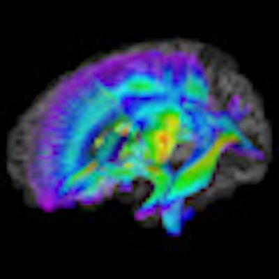
 |
| Image of white-matter pathways extracted from DTI-MRI for infants at risk of developing autism. Warmer colors represent greater fractional anisotropy values. Image courtesy of the University of North Carolina and the American Journal of Psychiatry. |
In addition, infants who developed autism had elevated fractional anisotropy values at six months, but then experienced slower change over time. By 24 months, infants with autism had lower fractional anisotropy values compared to infants without autism.
"These results suggest that aberrant development of white-matter pathways may precede the manifestation of autism symptoms in the first year of life," the authors concluded.
Wolff and colleagues also noted that the presence of "significant differences in fractional anisotropy at six months raises the exciting possibility of developing imaging biomarkers for risk of ASDs in advance of symptom onset. Future work might investigate the potential of predictive models for ASDs in early infancy, a process that could include refined imaging techniques or combined biobehavioral markers," they wrote.
Identifying high-risk infants before autism manifests itself "offers the possibility of implementing interventions that could reduce or even prevent the manifestation of the full syndrome," they added.
The results are the latest findings from the ongoing Infant Brain Imaging Study (IBIS) Network, which is funded by the U.S. National Institutes of Health and is headquartered at the University of North Carolina.