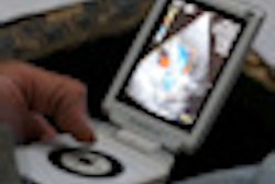
A handheld ultrasound system used for point-of-care heart studies offers accurate assessment of major heart functions, raising the potential for the systems to be used in place of more expensive traditional echocardiography scanners, according to research published in the July 5 edition of Annals of Internal Medicine.
A research project led by Scripps Translational Science Institute and Scripps Health found that the handheld echo system provided accurate assessments of ejection fraction and other measures for assessing heart health in patients. As a result, the authors, led by Dr. Eric Topol, believe the technology could help reduce unnecessary echocardiograms, particularly when used by a clinician trained in obtaining and interpreting the images.
The researchers compared the Vscan handheld ultrasound system (GE Healthcare) with transthoracic echocardiography (TTE) in a convenience sample of 97 consecutive patients referred for echocardiography. Scans were obtained from Vscan to visually assess various heart structures within a five-minute time frame to simulate the length of time of a physical examination, according to the researchers (Ann Intern Med, July 5, 2011, Vol. 155:1, pp. 33-38).
The study team then compared conclusions drawn from these imaging results with conclusions obtained from imaging with standard TTE systems. The researchers also evaluated the ability of observers to visualize heart structures, as well as their accuracy in image interpretation.
In addition, the group examined accuracy differences between cardiology attending physicians and less-experienced cardiology fellows who had two or fewer months of experience in echocardiographic interpretation.
The adequacy of handheld ultrasound images for visualization was rated as follows:
- Ejection fraction: 95%
- Wall-motion abnormality: 83%
- Left ventricular end-diastolic dimension: 95%
- Pericardial effusion: 94%
- Mitral valve: 90%
- Aortic valve: 82%
- Inferior vena cava size: 75%
As for interpretation accuracy, the handheld ultrasound images were most accurate for assessing ejection fractions and aortic valves, and they were least accurate for assessing inferior vena cava size, according to the study team. In addition, the authors noted higher accuracy and between-physician agreement for the experienced cardiologists than with the less-experienced cardiology follows.