It's not up for an Academy Award, but a new action movie starring a painful wrist could help doctors detect subtle dynamic joint instabilities that can be seen only in motion.
Researchers from the Mayo Clinic in Rochester, MN, showed how nongated 4D CT scans offer a way to diagnose dynamic joint instabilities at an early stage, allowing early intervention that can prevent further deterioration.
"Osteoarthritis is one of the most pervasive diseases," said Shuai Leng, PhD, in a presentation at the 2010 RSNA meeting in Chicago. "Patients suffering from joint disability experience progressive degenerative disease that results in functional instability osteoarthritis" or static deformities, he said.
"Early intervention can restore normal function before the onset of arthritis, so there's a big need to diagnose joint instability," Leng continued.


Few alternatives
For many join instability cases, there are few alternatives for diagnosis. Joint instability can be classified as static or dynamic, Leng explained, and the dynamic type can only be detected through joint motion, he said. Therefore, the study aimed to develop a high temporal and spatial resolution 4D CT technique for evaluating dynamic joint instability.
"Previously there were some studies that used ... retrospectively [electrocardiogram (ECG)-gated] cardiac CT, and in this technique multiple cycles are needed and periodic motion is required, which might be difficult for patients to maintain," Leng said.
The new exam is very similar to a CT perfusion study, he said. "There's no table motion and it only requires a single motion cycle, so it doesn't require periodic motion."
The dynamic wrist scan study was first performed on a human cadaver forearm.
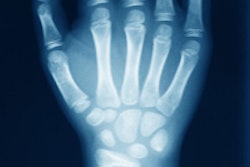
All images were acquired in sequential cardiac scan mode on a dual-source CT scanner (Somatom Definition Flash, Siemens Healthcare). The nongated perfusionlike scan mode was used to acquire continuous projection data from each x-ray source for two seconds, the duration of a single-motion cycle. The z-axis coverage was 38.4 mm, large enough to fully encompass the proximal carpal bones.
Images were reconstructed at 20 different time points using a commercial dual-source cardiac reconstruction algorithm, which provides 75-msec temporal resolution, sufficient to capture the moving wrist. A volume-rendering technique was used to generate 3D images, and a 4D movie was generated from 3D images at all 20 time points.
The wrist was scanned before and after cutting all three portions of the scapholunate ligament (simulating scapholunate instability). Viewing the movie with the intact ligament, the scaphoid and lunate move together, but once the ligament is cut it's easy to see that they move separately, demonstrating the ability of the 4D CT technique to detect dynamic joint instability.
"We showed this result to three orthopedic surgeons and we asked them to blindly review the images to tell us which, if any, had joint instability and they identified them almost instantaneously -- they didn't even need to see the whole movie," he said.
The still images were also useful, Leng said. Carpal bones, distal radius and ulna, and joint spaces were clearly delineated in the reconstructed 2D images and 3D images, and there was no motion blurring or banding artifacts. A hand surgeon judged the images to be of excellent quality.
 |
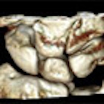
| Volume-rendered images of cadaver wrist in radial deviation, neutral, and ulna deviation positions show individual carpal bones and joint spaces clearly in three dimensions. All images courtesy of Shuai Leng, PhD. |
To reduce the radiation dose, the group rescanned the wrist at several levels, and was able to achieve diagnostic image quality at 120 kV and 50 mAs, corresponding to a CTDIvol radiation dose of 18 mGy. Even without dose reduction, however, "the radiation dose is much lower than indicated for any stochastic effect," Leng said.
Finally, the experiment was repeated in a living human subject with similar excellent results, Leng reported.
"The 4D CT technique produces high temporal and spatial resolution images of the wrist joint," he said. "In this study, we were able to get temporal resolution of 75 msec, and joint instability can be detected."
Because the technique doesn't require periodic motion on the part of the subject, it provides a more robust exam than earlier retrospectively gated techniques, and it may allow diagnosis of dynamic joint instabilities that can only be detected during joint motion, he added.
One drawback to using the technique to image other joints is detector size, which is only 4 cm for this scanner, but sufficient for a small joint like the wrist, Leng said.
Nongated 4D CT offers a way to diagnose dynamic joint instabilities at an early stage, enabling intervention to restore normal function before the onset of arthritis or static deformities, he concluded.
By Eric Barnes
AuntMinnie.com staff writer
February 22, 2011