MRI may be a more practical clinical biomarker for early detection of Alzheimer's disease than FDG-PET, according to a new study published in the September issue of Radiology.
Researchers from the University of California, San Diego (UCSD) in La Jolla found no evidence that FDG-PET is more sensitive than MRI to degeneration, which occurs in preclinical and mild cases of Alzheimer's disease (Radiology, 2010, Vol. 256:3, pp. 932-942).
"FDG-PET measures were largely redundant with MR imaging measures at all stages of disease, with hippocampal volume exhibiting the overall largest effect," wrote lead author David Karow, MD, PhD, and his colleagues from UCSD's departments of radiology, neuroscience, and psychiatry. "These results suggest that MR imaging measures, such as hippocampal volume, are at least as robust as FDG-PET measures even at early preclinical stages and may be more practical for clinical use in early detection of [Alzheimer's disease]."
The researchers gathered patient information from the Alzheimer's Disease Neuroimaging Initiative (ADNI), analyzing MRI and FDG-PET data between May 2008 and clinical follow-up through March 2010.
Enrollment criteria
Enrolled patients ranged in age from 55 to 90 years and were in good general health. The study excluded patients in a skilled nursing facility, individuals with any neurologic diseases other than Alzheimer's or those who were suspected of Alzheimer's disease, and patients with MR images that indicated infection, infarction, or other focal lesions.
The final study population included 80 healthy control subjects, 68 patients with Alzheimer's disease, and 156 individuals with amnestic mild cognitive impairment, of whom 69 had single-domain amnestic mild cognitive impairment.
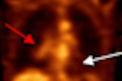
The researchers also downloaded dual 3D T1-weighted MR images from the ADNI database and collected studies performed with a variety of 1.5-tesla MRI systems. FDG-PET data were collected as multiple frames of 3D data, with a total image time of 30 minutes, starting approximately 30 minutes after the injection of 5 mCi of FDG.
Regions of interest were determined after coregistering FDG-PET and MR images for each subcortical and cortical area.
 |
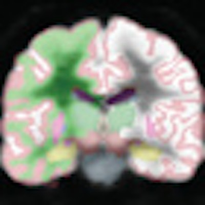
| Automated segmentation in a 71-year-old healthy male. MR image (left) segmentation is compared to segmentation of coregistered FDG-PET volume (right), with regions of interest including the hippocampus (gold), thalamus (dark green), caudate (blue), putamen (pink), and lateral ventricle (purple). The right and left hemisphere white matter (green and white, respectively) and gray matter (maroon) also are shown. Image courtesy of Radiology. |
With images in hand, the researchers compared results between the Alzheimer's disease group, the healthy control group, and the group with mild cognitive impairment.
Alzheimer's comparison
In the comparison between patients with Alzheimer's disease and the healthy controls, the researchers found widespread atrophy in cortical areas, with the greatest amount of atrophy in the hippocampus, which is responsible for learning and memory, and the entorhinal cortex, which can help measure the early stages of Alzheimer's disease.
The comparison also showed regional patterns of metabolic reductions overlapped with areas of atrophy, with the largest metabolic reductions observed in the mesial and lateral temporal, temporoparietal, and posterior cingulate regions. All three regions of the brain are known to be the first areas affected by Alzheimer's disease.
In addition, the volume of the hippocampus showed the single largest effect size of all structural measures and was significantly greater than the largest metabolic effect size, which was in the entorhinal cortex.
Mild cognitive impairment analysis
In the analysis of the group with mild cognitive impairment compared to the healthy control group, structural and metabolic reductions were seen in several regions of interest, but they were not as widespread as in the comparison between the Alzheimer's patients and the healthy controls.
Once again, the largest morphometric reductions were seen in the hippocampus and entorhinal cortex. Both structures also exhibited the largest reductions compared to results in the healthy control subjects.
In contrast, the authors noted, the posterior cingulate "exhibited significant metabolic reduction without significant concomitant changes in atrophy."
Comparing the group with mild cognitive impairment to the healthy control patients also revealed more focal structural and metabolic reductions. Significant atrophy was discovered in the mesial temporal regions only, with the largest reduction in the hippocampus. Significant metabolic reductions also were observed in the mesial temporal regions of interest and posterior cingulate, with the largest effect occurring for the entorhinal cortex.
Hippocampal volume
The analysis of follow-up data found that reduced hippocampal volume and reduced entorhinal metabolism were "each associated with significantly higher odds of conversion to Alzheimer's disease within two years," the authors wrote. "Although the odds ratio for hippocampal volume was higher than that for entorhinal metabolism, the overlap in [confidence interval] suggests that the two measures do not significantly differ."
Based on the results, Karow and colleagues concluded that FDG-PET measures were "largely redundant with MR imaging measures at all stages of disease, with hippocampal volume exhibiting the overall largest effect."
"These results suggest that MR imaging measures, such as hippocampal volume, are at least as robust as FDG-PET measures even at early preclinical stages and may be more practical for clinical use in early detection of [Alzheimer's disease]," they wrote.
By Wayne Forrest
AuntMinnie.com staff writer
August 27, 2010
Related Reading
FDG-PET and memory tests best predict Alzheimer's, June 30, 2010
MRI finds genetic link in Alzheimer's disease, June 14, 2010
PET can help clarify dementia diagnoses, May 3, 2010
MRI hippocampal measurement doesn't predict cognitive decline, April 26, 2010
MR scans may detect signs of early dementia in elderly, April 14, 2010
Copyright © 2010 AuntMinnie.com