Digital breast tomosynthesis (DBT) has the potential to be a useful tool for screening women with dense breasts. But the fact that it adds more radiation exposure is problematic, especially when compared to MRI, according to Dr. Stamatia Destounis from the Elizabeth Wende Breast Care Center (EWBC) in Rochester, NY.
Destounis offered an overview of the technology and its potential role in breast screening at this week's National Consortium of Breast Centers (NCBC) conference in Las Vegas.
Mammography is a 2D modality used to image a 3D structure, and issues such as patient motion and overlapped tissue can get in the way of accurate images. One benefit of digital breast tomosynthesis is that it combines 2D imaging with 3D imaging.
"The technology is different from a standard mammogram in the same way a CT of the chest is different from a standard chest x-ray," Destounis said. "One is three dimensional; the other is flat."
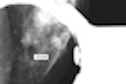
Digital breast tomosynthesis is performed by an x-ray tube moving through a proscribed arc. Fifteen low-dose projection images are acquired during a five-second sweep; during acquisition, the x-ray tube moves at ± 7.5°. Projections are then reconstructed into a series of images at 1-mm intervals. These slices can be viewed as a cine loop or as individual slices. DBT is currently being used for research only in the U.S., according to Destounis.
"The whole point is that with DBT you are able to reduce the overlap, and can improve screening and diagnostic accuracy," she told AuntMinnie.com.
Using a prototype unit from Hologic of Bedford, MA, EWBC has been conducting a study that compares DBT to conventional mammography for the characterization and visualization of breast calcifications. All patients will undergo a bilateral tomosynthesis exam, Destounis said.
Currently, the study has enrolled 52 patients, with an average patient age of 58 years. Of the 52 women, one has fatty breast tissue, 16 have scattered breast tissue, 23 have heterogeneously dense tissue, and 12 have dense tissue. Of these 52 patients, 15 malignant results were found, one atypical result was found, and 36 benign results were found. The average lesion size found on DBT is 16 mm, while the average lesion size found on 2D mammography is 13 mm, Destounis said.
What is DBT's future?
Destounis sees DBT as a potentially good screening tool for women with dense breasts. But because of concerns about radiation exposure, the technology may have a steep climb to clinical acceptance, especially since breast MRI has also been shown to work well as a screening tool for this population.
"DBT has a bit of an uphill battle," she told AuntMinnie.com. "MRI already has a niche for high-risk patients, and it doesn't use radiation. The tomosynthesis images are good: They show microcalcifications and the edges of masses well. But it's unclear whether tomosynthesis can stand alone as a screening tool for high-risk women, and if it can't, adding it to standard mammography will certainly increase the radiation exposure a woman receives."
By Kate Madden Yee
AuntMinnie.com staff writer
March 22, 2010
Related Reading
Adding DBT to mammo increases cancer detection in dense breasts, January 18, 2010
DBT reduces false-positive rate by 41% in screening setting, December 1, 2009
Digital breast tomosynthesis cuts recall rate by 30%, August 4, 2009
ECR delivers new findings on digital breast tomosynthesis, March 7, 2009
New breast imaging applications show diagnostic promise, January 20, 2009
Copyright © 2010 AuntMinnie.com